5
views
0
recommends
+1 Recommend
0 collections
    0
    shares
      • Record: found
      • Abstract: found
      • Article: found
      Is Open Access

      Synergistic enzymatic and bioorthogonal reactions for selective prodrug activation in living systems

      research-article

      Read this article at

      Bookmark
          There is no author summary for this article yet. Authors can add summaries to their articles on ScienceOpen to make them more accessible to a non-specialist audience.

          Abstract

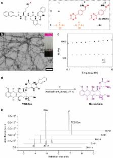
          Adverse drug reactions (ADRs) restrict the maximum doses applicable in chemotherapy, which leads to failure in cancer treatment. Various approaches, including nano-drug and prodrug strategies aimed at reducing ADRs, have been developed, but these strategies have their own pitfalls. A renovated strategy for ADR reduction is urgently needed. Here, we employ an enzymatic supramolecular self-assembly process to accumulate a bioorthogonal decaging reaction trigger inside targeted cancer cells, enabling spatiotemporally controlled, synergistic prodrug activation. The bioorthogonally activated prodrug exhibits significantly enhanced potency against cancer cells compared with normal cells. This prodrug activation strategy further demonstrates high tumour inhibition efficacy with satisfactory biocompatibility, pharmacokinetics, and safety in vivo. We envision that integration of enzymatic and bioorthogonal reactions will serve as a general small-molecule-based strategy for alleviation of ADRs in chemotherapy.

          Abstract

          The side effects of cancer drugs limit their utility. Here, the authors developed a method in which an inactive (prodrug) version of the cancer drug doxorubicin enters tumour cells and then gets activated inside the cells upon a trigger facilitated by enzyme-instructed supramolecular self-assembly.

          Related collections

          Most cited references29

          • Record: found
          • Abstract: found
          • Article: not found

          Tumor vascular permeability, accumulation, and penetration of macromolecular drug carriers.

          Delivery of anticancer therapeutic agents to solid tumors is problematic. Macromolecular drug carriers are an attractive alternative drug delivery method because they appear to target tumors and have limited toxicity in normal tissues. We investigated how molecular weight influences the accumulation of a model macromolecular drug carrier, dextran covalently linked to a fluorophore, in tumors. We used dextrans with molecular weights from 3.3 kDa to 2 MDa. Vascular permeability, accumulation, and three-dimensional penetration of these dextrans were simultaneously measured in solid tumors via a dorsal skin fold window chamber, intravital laser-scanning confocal microscopy, and custom image analysis. Increasing the molecular weight of dextran statistically significantly reduced its vascular permeability by approximately two orders of magnitude (i.e., from 154 x 10(-7) cm/s, 95% confidence interval [CI] = 134 to 174 x 10(-7) cm/s, for 3.3-kDa dextran to 1.7 x 10(-7) cm/s, 95% CI = 0.7 to 2.6 x 10(-7) cm/s for 2-MDa dextran; P < .001, two-sided Kruskal-Wallis test) but increased its plasma half-life, which provided ample time for extravasation (i.e., to enter tumor tissue from the vasculature). Tumor accumulation was maximal for dextrans with molecular weights between 40 and 70 kDa. Dextrans of 3.3 and 10 kDa penetrated deeply (greater than 35 microm) and homogeneously into tumor tissue from the vessel wall. After a 30-minute period, a high concentration was observed only approximately 15 microm from the vessel wall for 40- to 70-kDa dextrans and only 5 microm for 2-MDa dextrans. Increasing the molecular weight of dextran statistically significantly reduced its tumor vascular permeability. Dextrans of 40 and 70 kDa had the highest accumulation in solid tumors but were largely concentrated near the vascular surface.
            Bookmark
            • Record: found
            • Abstract: found
            • Article: not found

            Bioorthogonal Cyclization-Mediated In Situ Self-Assembly of Small Molecule Probes for Imaging Caspase Activity in vivo

            Directed self-assembly of small molecules in living systems could enable a myriad of applications in biology and medicine, and it has been widely used to synthesize supramolecules and nano/microstructures in solution and in living cells. However, controlling self-assembly of synthetic small molecules in living animals is challenging because of the complex and dynamic in vivo physiological environment. Here we employed an optimized first-order bioorthogonal cyclization reaction to control self-assembly of a fluorescent small molecule, and demonstrated its in vivo applicability by imaging of casapae-3/7 activity in human tumor xenograft mouse models of chemotherapy. The in situ assembled fluorescent nanoparticles have been successfully imaged in both apoptotic cells and tumor tissues using three-dimensional structured illumination microscopy. This strategy combines the advantages offered by small molecules with those of nanomaterials and should find widespread use for non-invasive imaging of enzyme activity in vivo.
              Bookmark
              • Record: found
              • Abstract: found
              • Article: not found

              Imaging enzyme-triggered self-assembly of small molecules inside live cells

              Self-assembly of small molecules in water to form nanofibers, besides generating sophisticated biomaterials, promises a simple system inside cells for regulating cellular processes. But lack of a convenient approach for studying the self-assembly of small molecules inside cells hinders the development of such systems. Here we report a method to image enzyme-triggered self-assembly of small molecules inside live cells. After linking a fluorophore to a self-assembly motif to make a precursor, we confirmed by 31P NMR and rheology that enzyme-triggered conversion of the precursor to a hydrogelator results in the formation of a hydrogel via self-assembly. The imaging contrast conferred by the nanofibers of the hydrogelators allowed the evaluation of intracellular self-assembly; the dynamics, and the localization of the nanofibers of the hydrogelators in live cells. This approach explores supramolecular chemistry inside cells and may lead to new insights, processes, or materials at the interface of chemistry and biology.
                Bookmark

                Author and article information

                Contributors
                pengchen@pku.edu.cn
                gaoy@nanoctr.cn
                Journal
                Nat Commun
                Nat Commun
                Nature Communications
                Nature Publishing Group UK (London )
                2041-1723
                28 November 2018
                28 November 2018
                2018
                : 9
                : 5032
                Affiliations
                [1 ]ISNI 0000 0004 1806 6075, GRID grid.419265.d, CAS Center for Excellence in Nanoscience, CAS Key Laboratory of Biomedical Effects of Nanomaterials and Nanosafety, , National Center for Nanoscience and Technology, ; Beijing, 100190 China
                [2 ]ISNI 0000 0004 1797 8419, GRID grid.410726.6, University of Chinese Academy of Sciences, ; Beijing, 100049 China
                [3 ]ISNI 0000 0001 2256 9319, GRID grid.11135.37, Key Laboratory of Bioorganic Chemistry and Molecular Engineering of Ministry of Education, College of Chemistry and Molecular Engineering, , Peking University, ; Beijing, 100871 China
                [4 ]ISNI 0000 0001 2256 9319, GRID grid.11135.37, Peking-Tsinghua Center for Life Sciences, , Peking University, ; Beijing, 100871 China
                [5 ]ISNI 0000 0001 2256 9319, GRID grid.11135.37, Medical Isotopes Research Center and Department of Radiation Medicine, School of Basic Medical Sciences, , Peking University, ; Beijing, 100191 China
                Author information
                http://orcid.org/0000-0002-3099-8495
                http://orcid.org/0000-0001-8718-8154
                http://orcid.org/0000-0002-5008-4703
                http://orcid.org/0000-0002-0402-7417
                http://orcid.org/0000-0001-9714-4219
                Article
                7490
                10.1038/s41467-018-07490-6
                6261997
                30487642
                44d8c82f-f95b-412b-8254-8357c55006f1
                © The Author(s) 2018

                Open Access This article is licensed under a Creative Commons Attribution 4.0 International License, which permits use, sharing, adaptation, distribution and reproduction in any medium or format, as long as you give appropriate credit to the original author(s) and the source, provide a link to the Creative Commons license, and indicate if changes were made. The images or other third party material in this article are included in the article’s Creative Commons license, unless indicated otherwise in a credit line to the material. If material is not included in the article’s Creative Commons license and your intended use is not permitted by statutory regulation or exceeds the permitted use, you will need to obtain permission directly from the copyright holder. To view a copy of this license, visit http://creativecommons.org/licenses/by/4.0/.

                History
                : 20 May 2018
                : 6 November 2018
                Categories
                Article
                Custom metadata
                © The Author(s) 2018

                Uncategorized
                Uncategorized

                Comments

                Comment on this article