31
views
0
recommends
+1 Recommend
0 collections
    0
    shares
      • Record: found
      • Abstract: found
      • Article: found
      Is Open Access

      JAK-STAT and G-protein-coupled receptor signaling pathways are frequently altered in epitheliotropic intestinal T-cell lymphoma

      research-article
      1 , 1 , 2 , 3 , 1 , 2 , 3 , 2 , 3 , 2 , 3 , 2 , 3 , 1 , 1 , 2 , 3 , 2 , 3 , 3 , 4 , 1 , 2 , 3 , 1 , 1 , 1 , 5 , 6 , 7 , 8 , 8 , 9 , 10 , 10 , 5 , 11 , 12 , 13 , 14 , 15 , 15 , 16 , 17 , 18 , 19 , 5 , 20 , 21 , 21 , 21 , 21 , 21 , 3 , 4 , 3 , 5 , 22 , 2 , 3 , 5 , 23 , 1 , 21 , 24 , * , 7 , 11 , 12 , 15 , 18 , 23 , * , 1 , *
      Leukemia
      Nature Publishing Group

      Read this article at

      Bookmark
          There is no author summary for this article yet. Authors can add summaries to their articles on ScienceOpen to make them more accessible to a non-specialist audience.

          Abstract

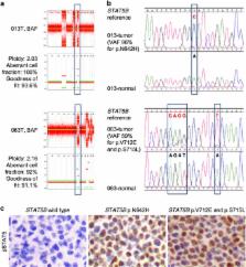
          Epitheliotropic intestinal T-cell lymphoma (EITL, also known as type II enteropathy-associated T-cell lymphoma) is an aggressive intestinal disease with poor prognosis and its molecular alterations have not been comprehensively characterized. We aimed to identify actionable easy-to-screen alterations that would allow better diagnostics and/or treatment of this deadly disease. By performing whole-exome sequencing of four EITL tumor-normal pairs, followed by amplicon deep sequencing of 42 tumor samples, frequent alterations of the JAK-STAT and G-protein-coupled receptor (GPCR) signaling pathways were discovered in a large portion of samples. Specifically, STAT5B was mutated in a remarkable 63% of cases, JAK3 in 35% and GNAI2 in 24%, with the majority occurring at known activating hotspots in key functional domains. Moreover, STAT5B locus carried copy-neutral loss of heterozygosity resulting in the duplication of the mutant copy, suggesting the importance of mutant STAT5B dosage for the development of EITL. Dysregulation of the JAK-STAT and GPCR pathways was also supported by gene expression profiling and further verified in patient tumor samples. In vitro overexpression of GNAI2 mutants led to the upregulation of pERK1/2, a member of MEK-ERK pathway. Notably, inhibitors of both JAK-STAT and MEK-ERK pathways effectively reduced viability of patient-derived primary EITL cells, indicating potential therapeutic strategies for this neoplasm with no effective treatment currently available.

          Related collections

          Most cited references35

          • Record: found
          • Abstract: found
          • Article: not found

          Patterns of somatic mutation in human cancer genomes.

          Cancers arise owing to mutations in a subset of genes that confer growth advantage. The availability of the human genome sequence led us to propose that systematic resequencing of cancer genomes for mutations would lead to the discovery of many additional cancer genes. Here we report more than 1,000 somatic mutations found in 274 megabases (Mb) of DNA corresponding to the coding exons of 518 protein kinase genes in 210 diverse human cancers. There was substantial variation in the number and pattern of mutations in individual cancers reflecting different exposures, DNA repair defects and cellular origins. Most somatic mutations are likely to be 'passengers' that do not contribute to oncogenesis. However, there was evidence for 'driver' mutations contributing to the development of the cancers studied in approximately 120 genes. Systematic sequencing of cancer genomes therefore reveals the evolutionary diversity of cancers and implicates a larger repertoire of cancer genes than previously anticipated.
            Bookmark
            • Record: found
            • Abstract: found
            • Article: not found

            Integration of gene mutations in risk prognostication for patients receiving first-line immunochemotherapy for follicular lymphoma: a retrospective analysis of a prospective clinical trial and validation in a population-based registry.

            Follicular lymphoma is a clinically and genetically heterogeneous disease, but the prognostic value of somatic mutations has not been systematically assessed. We aimed to improve risk stratification of patients receiving first-line immunochemotherapy by integrating gene mutations into a prognostic model.
              Bookmark
              • Record: found
              • Abstract: found
              • Article: not found

              The emerging mutational landscape of G proteins and G-protein-coupled receptors in cancer.

              Aberrant expression and activity of G proteins and G-protein-coupled receptors (GPCRs) are frequently associated with tumorigenesis. Deep sequencing studies show that 4.2% of tumours carry activating mutations in GNAS (encoding Gαs), and that oncogenic activating mutations in genes encoding Gαq family members (GNAQ or GNA11) are present in ~66% and ~6% of melanomas arising in the eye and skin, respectively. Furthermore, nearly 20% of human tumours harbour mutations in GPCRs. Many human cancer-associated viruses also express constitutively active viral GPCRs. These studies indicate that G proteins, GPCRs and their linked signalling circuitry represent novel therapeutic targets for cancer prevention and treatment.
                Bookmark

                Author and article information

                Journal
                Leukemia
                Leukemia
                Leukemia
                Nature Publishing Group
                0887-6924
                1476-5551
                June 2016
                08 February 2016
                01 March 2016
                : 30
                : 6
                : 1311-1319
                Affiliations
                [1 ]Lymphoma Genomic Translational Research Laboratory, Division of Medical Oncology, National Cancer Centre Singapore , Singapore, Singapore
                [2 ]Laboratory of Cancer Epigenome, Division of Medical Sciences, National Cancer Centre Singapore , Singapore, Singapore
                [3 ]Program in Cancer and Stem Cell Biology, Duke-NUS Medical School , Singapore, Singapore
                [4 ]Centre for Computational Biology, Duke-NUS Medical School , Singapore, Singapore
                [5 ]Cancer Science Institute of Singapore, National University of Singapore , Singapore, Singapore
                [6 ]Department of Biomedical Engineering, Duke University , Durham, NC, USA
                [7 ]Department of Pathology, Singapore General Hospital , Singapore, Singapore
                [8 ]Advanced Molecular Pathology Laboratory, Singapore Health Services , Singapore, Singapore
                [9 ]Department of Pathology, Sun Yat-sen University Cancer Center , Guangzhou, China
                [10 ]Department of Pathology, Tan Tock Seng Hospital , Singapore, Singapore
                [11 ]Department of Pathology, Yong Loo Lin School of Medicine, National University of Singapore , Singapore, Singapore
                [12 ]Department of Pathology, National University Hospital, National University Health System , Singapore, Singapore
                [13 ]Department of Pathology, Chi Mei Medical Center , Tainan, Taiwan
                [14 ]Department of Pathology, Taipei Medical University and National Taiwan University , Taipei, Taiwan
                [15 ]Department of Pathology, Guangdong General Hospital , Guangzhou, China
                [16 ]Department of Pathology, Faculty of Medicine Siriraj Hospital, Mahidol University , Bangkok, Thailand
                [17 ]Department of Pathology, Samsung Medical Centre, Sungkyunkwan University School of Medicine , Seoul, South Korea
                [18 ]Department of Pathology, University of Malaya , Kuala Lumpur, Malaysia
                [19 ]Department of Pathology, Hospital Raja Permaisuri Bainun , Ipoh, Malaysia
                [20 ]Department of Haematology-Oncology, National University Hospital, National University Health System , Singapore, Singapore
                [21 ]Division of Medical Oncology, National Cancer Centre Singapore , Singapore, Singapore
                [22 ]Genome Institute of Singapore, A*STAR , Singapore, Singapore
                [23 ]Institute of Molecular and Cell Biology, A*STAR , Singapore, Singapore
                [24 ]Office of Education, Duke-NUS Medical School , Singapore, Singapore
                Author notes
                [* ]Lymphoma Genomic Translational Research Laboratory, Division of Medical Oncology, National Cancer Centre Singapore , 11 Hospital Drive, Singapore 169610, Singapore E-mail: dmolst@ 123456nccs.com.sg or cmrock@ 123456nccs.com.sg .
                [* ]Department of Pathology, Singapore General Hospital , 20 College Road, Singapore 169856, Singapore. E-mail: drtansy@ 123456gmail.com
                [25]

                These authors contributed equally to this work.

                Article
                leu201613
                10.1038/leu.2016.13
                4895162
                26854024
                45c629ed-32d7-4b52-9d18-7ae6f6630dd6
                Copyright © 2016 Macmillan Publishers Limited

                This work is licensed under a Creative Commons Attribution-NonCommercial-NoDerivs 4.0 International License. The images or other third party material in this article are included in the article's Creative Commons license, unless indicated otherwise in the credit line; if the material is not included under the Creative Commons license, users will need to obtain permission from the license holder to reproduce the material. To view a copy of this license, visit http://creativecommons.org/licenses/by-nc-nd/4.0/

                History
                : 15 July 2015
                : 07 January 2016
                : 18 January 2016
                Categories
                Original Article

                Oncology & Radiotherapy
                Oncology & Radiotherapy

                Comments

                Comment on this article