21
views
0
recommends
+1 Recommend
0 collections
    0
    shares
      • Record: found
      • Abstract: found
      • Article: found
      Is Open Access

      Utility of Cardiac Magnetic Resonance in the Evaluation of Unselected Patients with Possible Arrhythmogenic Right Ventricular Cardiomyopathy

      research-article

      Read this article at

      Bookmark
          There is no author summary for this article yet. Authors can add summaries to their articles on ScienceOpen to make them more accessible to a non-specialist audience.

          Abstract

          Introduction:

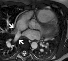
          Arrhythmogenic right ventricular cardiomyopathy (ARVC) is a rare but important cause of sudden cardiac death. We investigated the role of cardiac magnetic resonance imaging (CMR) in the evaluation of patients with suspected ARVC referred by a general cardiology service.

          Methods:

          Ninety-two patients (mean age 48 ± 15, 49% female), referred for CMR assessment of possible ARVC, were reviewed. CMR included both functional and tissue characteristic imaging.

          Results:

          No patients had ARVC based on the 1994 Task Force Criteria (TFC) prior to CMR, but 4 met proposed Modified TFC; 15% met one major (±1 minor) TFC, 71% 1 or 2 minor TFC, and 14% no TFC. Reasons for CMR referral included symptomatic arrhythmia of likely RV origin (28%), Electrocardiogram/Holter abnormalities (28%), echocardiographic features suspicious of ARVC (19%), and family history of ARVC (8%). CMR findings strongly suggestive of ARVC were found in nine patients (10%), although only three were considered typical. Of these patients two met 1 major TFC and seven met 1 or 2 minor TFC. CMR findings included RV thinning, aneurysm, and diastolic out-pouching, but only 1 patient had definite fatty infiltration of the RV. Incidentally, CMR detected important, previously undiagnosed pathology, including anomalous pulmonary venous drainage (2 patients) and non-ischaemic cardiomyopathy (6%). CMR was normal in 63%, with minor abnormalities in 29%.

          Conclusions:

          CMR may play an important diagnostic role in the evaluation of possible ARVC. Patients who do not meet TFC for diagnosis may have CMR features typical of ARVC. Additionally CMR may detect other hitherto undiagnosed structural or functional abnormalities that alter patient management. However the majority of patients referred have a low pretest probability of ARVC, and the rate of normal CMR scans is high.

          Related collections

          Most cited references20

          • Record: found
          • Abstract: not found
          • Article: not found

          Diagnosis of arrhythmogenic right ventricular dysplasia/cardiomyopathy. Task Force of the Working Group Myocardial and Pericardial Disease of the European Society of Cardiology and of the Scientific Council on Cardiomyopathies of the International Society and Federation of Cardiology.

            Bookmark
            • Record: found
            • Abstract: found
            • Article: not found

            Normal human left and right ventricular and left atrial dimensions using steady state free precession magnetic resonance imaging.

            The aim of this project was to establish a database of left and right ventricular and left atrial dimensions in healthy volunteers using steady-state free precession cardiac magnetic resonance imaging, the clinical technique of choice, across a wide age range. 108 healthy volunteers (63 male, 45 female) underwent cardiac magnetic resonance imaging using steady-state free precession sequences. Manual analysis was performed by 2 experienced observers. Left and right ventricular volumes and left ventricular mass were larger in males than females: LV end-diastolic volume 160 +/- 29 mL vs. 135 +/- 26 mL, LV end-systolic volume 50 +/- 16 mL vs. 42 +/- 12 mL; RV end-diastolic volume 190 +/- 33 mL vs. 148 +/- 35 mL, RV end-systolic volume 78 +/- 20 mL vs. 56 +/- 18 mL (p < .05 for all). Normalization of values to body surface area removed the statistical differences for LV volumes, but not for LV mass or RV volumes. With increased age, males showed a significant decrease in volume and mass indices for both ventricles, while female values remained unchanged. Compared to females, males had significantly larger maximal left atrial volumes (103 +/- 30 mL vs. 89 +/- 21 mL, p = .01) and left atrial stroke volumes (58 +/- 23 mL vs. 48 +/- 15 mL, p = .01). There was no difference in left atrial ejection fraction between the sexes. We have produced a large database of age-related normal ranges for left and right ventricular function and left atrial function in males and females. This will allow accurate interpretation of clinical and research datasets.
              Bookmark
              • Record: found
              • Abstract: found
              • Article: not found

              Contrast enhanced-cardiovascular magnetic resonance imaging in patients with pulmonary hypertension.

              To determine the presence and extent of delayed contrast enhancement (DCE) in patients with pulmonary hypertension (PHT) using contrast enhanced-cardiovascular magnetic resonance imaging (ce-CMR). Twenty-five patients with PHT underwent ce-CMR and right heart catheterization. Right ventricular (RV) and left ventricular (LV) volumes, ejection fraction, mass, and DCE mass were determined with ce-CMR. Mean pulmonary artery pressure (mean PAP) averaged 43 (12) mmHg and cardiac output 4.3 (1.2) L/min. DCE was demonstrated in 23 out of 25 patients. DCE was confined to the RV insertion points (RVIPs) in seven patients and extended into the interventricular septum (IVS) in the remaining 16 patients. In these 16 patients, septal contrast enhancement was associated with IVS bowing. The extent of contrast enhancement correlated positively with RV end-diastolic volume/body surface area, RV mass, mean PAP, and pulmonary vascular resistance and correlated inversely with RV ejection fraction. DCE was present within the RVIPs and IVS of most patients with PHT studied. Extent of DCE correlated with RV function and pulmonary haemodynamics. DCE was associated with IVS bowing and may provide a novel marker for occult septal abnormalities directly relating to the haemodynamic stress experienced by these patients.
                Bookmark

                Author and article information

                Journal
                Clin Med Insights Cardiol
                Clin Med Insights Cardiol
                101466475
                Clinical Medicine Insights. Cardiology
                Libertas Academica
                1179-5468
                2012
                19 November 2012
                : 6
                : 153-162
                Affiliations
                Cardiovascular Division, North Shore Hospital, Waitemata Health.
                Author notes
                Corresponding author email: khangli@ 123456hotmail.com
                Article
                cmc-6-2012-153
                10.4137/CMC.S9996
                3511051
                23226076
                46d8c84d-366a-4623-a4a5-3d2f144af68f
                © 2012 the author(s), publisher and licensee Libertas Academica Ltd.

                This is an open access article. Unrestricted non-commercial use is permitted provided the original work is properly cited.

                History
                Categories
                Original Research

                Cardiovascular Medicine
                cardiac magnetic resonance imaging,cardiomyopathy
                Cardiovascular Medicine
                cardiac magnetic resonance imaging, cardiomyopathy

                Comments

                Comment on this article