23
views
0
recommends
+1 Recommend
0 collections
    0
    shares
      • Record: found
      • Abstract: found
      • Article: found
      Is Open Access

      Induction and transport of Wnt 5a during macrophage-induced malignant invasion is mediated by two types of extracellular vesicles

      research-article

      Read this article at

      Bookmark
          There is no author summary for this article yet. Authors can add summaries to their articles on ScienceOpen to make them more accessible to a non-specialist audience.

          Abstract

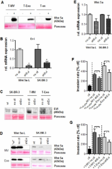
          Recently, we have shown that macrophage (MΦ)-induced invasion of breast cancer cells requires upregulation of Wnt 5a in MΦ leading to activation of β-Catenin-independent Wnt signaling in the tumor cells. However, it remained unclear, how malignant cells induce Wnt 5a in MΦ and how it is transferred back to the cancer cells. Here we identify two types of extracellular particles as essential for this intercellular interaction in both directions. Plasma membrane-derived microvesicles (MV) as well as exosomes from breast cancer cells, although biologically distinct populations, both induce Wnt 5a in MΦ. In contrast, the particle-free supernatant and vesicles from benign cells, such as platelets, have no such effect. Induction is antagonized by the Wnt inhibitor Dickkopf-1. Subsequently, Wnt 5a is shuttled via responding MΦ-MV and exosomes to the tumor cells enhancing their invasion. Wnt 5a export on both vesicle fractions depends at least partially on the cargo protein Evenness interrupted (Evi). Its knockdown leads to Wnt 5a depletion of both particle populations and reduced vesicle-mediated invasion. In conclusion, MV and exosomes are critical for MΦ-induced invasion of cancer cells since they are responsible for upregulation of MΦ-Wnt 5a as well as for its delivery to the recipient cells via a reciprocal loop. Although of different biogenesis, both populations share common features regarding function and Evi-dependent secretion of non-canonical Wnts.

          Related collections

          Most cited references19

          • Record: found
          • Abstract: found
          • Article: found
          Is Open Access

          Isolation and Characterization of RNA-Containing Exosomes

          The field of exosome research is rapidly expanding, with a dramatic increase in publications in recent years. These small vesicles (30-100 nm) of endocytic origin were first proposed to function as a way for reticulocytes to eradicate the transferrin receptor while maturing into erythrocytes1, and were later named exosomes. Exosomes are formed by inward budding of late endosomes, producing multivesicular bodies (MVBs), and are released into the environment by fusion of the MVBs with the plasma membrane2. Since the first discovery of exosomes, a wide range of cells have been shown to release these vesicles. Exosomes have also been detected in several biological fluids, including plasma, nasal lavage fluid, saliva and breast milk3-6. Furthermore, it has been demonstrated that the content and function of exosomes depends on the originating cell and the conditions under which they are produced. A variety of functions have been demonstrated for exosomes, such as induction of tolerance against allergen7,8, eradication of established tumors in mice9, inhibition and activation of natural killer cells10-12, promotion of differentiation into T regulatory cells13, stimulation of T cell proliferation14 and induction of T cell apoptosis15. Year 2007 we demonstrated that exosomes released from mast cells contain messenger RNA (mRNA) and microRNA (miRNA), and that the RNA can be shuttled from one cell to another via exosomes. In the recipient cells, the mRNA shuttled by exosomes was shown to be translated into protein, suggesting a regulatory function of the transferred RNA16. Further, we have also shown that exosomes derived from cells grown under oxidative stress can induce tolerance against further stress in recipient cells and thus suggest a biological function of the exosomal shuttle RNA17. Cell culture media and biological fluids contain a mixture of vesicles and shed fragments. A high quality isolation method for exosomes, followed by characterization and identification of the exosomes and their content, is therefore crucial to distinguish exosomes from other vesicles and particles. Here, we present a method for the isolation of exosomes from both cell culture medium and body fluids. This isolation method is based on repeated centrifugation and filtration steps, followed by a final ultracentrifugation step in which the exosomes are pelleted. Important methods to identify the exosomes and characterize the exosomal morphology and protein content are highlighted, including electron microscopy, flow cytometry and Western blot. The purification of the total exosomal RNA is based on spin column chromatography and the exosomal RNA yield and size distribution is analyzed using a Bioanalyzer.
            Bookmark
            • Record: found
            • Abstract: found
            • Article: not found

            Wnt5a signaling directly affects cell motility and invasion of metastatic melanoma.

            Gene expression profiling identified human melanoma cells demonstrating increased cell motility and invasiveness. The gene WNT5A best determined in vitro invasive behavior. Melanoma cells were transfected with vectors constitutively overexpressing Wnt5a. Consistent changes included actin reorganization and increased cell adhesion. No increase in beta-catenin expression or nuclear translocation was observed. There was, however, a dramatic increase in activated PKC. In direct correlation with Wnt5a expression and PKC activation, there was an increase in melanoma cell invasion. Blocking this pathway using antibodies to Frizzled-5, the receptor for Wnt5a, inhibited PKC activity and cellular invasion. Furthermore, Wnt5a expression in human melanoma biopsies directly correlated to increasing tumor grade. These observations support a role for Wnt5a in human melanoma progression.
              Bookmark
              • Record: found
              • Abstract: found
              • Article: not found

              Trans-synaptic transmission of vesicular Wnt signals through Evi/Wntless.

              Wnts play pivotal roles during development and in the mature nervous system. However, the mechanism by which Wnts traffic between cells has remained elusive. Here we demonstrate a mechanism of Wnt transmission through release of exosome-like vesicles containing the Wnt-binding protein Evenness Interrupted/Wntless/Sprinter (Evi/Wls/Srt). We show that at the Drosophila larval neuromuscular junction (NMJ), presynaptic vesicular release of Evi is required for the secretion of the Wnt, Wingless (Wg). We also show that Evi acts cell-autonomously in the postsynaptic Wnt-receiving cell to target dGRIP, a Wg-receptor-interacting protein, to postsynaptic sites. Upon Evi loss of function, dGRIP is not properly targeted to synaptic sites, interfering with postsynaptic Wnt signal transduction. These findings uncover a previously unknown cellular mechanism by which a secreted Wnt is transported across synapses by Evi-containing vesicles and reveal trafficking functions of Evi in both the Wnt-producing and the Wnt-receiving cells. For a video summary of this article, see the PaperFlick file with the Supplemental Data available online.
                Bookmark

                Author and article information

                Journal
                Oncotarget
                Oncotarget
                ImpactJ
                Oncotarget
                Impact Journals LLC
                1949-2553
                November 2013
                21 October 2013
                : 4
                : 11
                : 2057-2066
                Affiliations
                1 Dept of Hematology/Oncology, University Medicine, Göttingen, Germany
                2 Div. Signaling and Functional Genomics, German Cancer Research Center, Heidelberg, Germany
                3 Max Planck Institute for Biophysical Chemistry, Göttingen, Germany
                Author notes
                Correspondence to: Claudia Binder, cbinder@ 123456med.uni-goettingen.de
                Article
                10.18632/oncotarget.1336
                3875769
                24185202
                4712f350-a9b5-466f-bfb3-33e2c454c8cc
                Copyright: © 2013 Menck et al.

                This is an open-access article distributed under the terms of the Creative Commons Attribution License, which permits unrestricted use, distribution, and reproduction in any medium, provided the original author and source are credited

                History
                : 27 August 2013
                : 19 October 2013
                Categories
                Research Paper

                Oncology & Radiotherapy
                breast cancer,exosomes,evi,microvesicles,macrophages,wnt 5a
                Oncology & Radiotherapy
                breast cancer, exosomes, evi, microvesicles, macrophages, wnt 5a

                Comments

                Comment on this article