12
views
0
recommends
+1 Recommend
0 collections
    0
    shares
      • Record: found
      • Abstract: found
      • Article: not found

      The Tölz Temporal Topography Study: Mapping the visual field across the life span. Part I: The topography of light detection and temporal-information processing

      research-article

      Read this article at

      ScienceOpenPublisherPMC
      Bookmark
          There is no author summary for this article yet. Authors can add summaries to their articles on ScienceOpen to make them more accessible to a non-specialist audience.

          Abstract

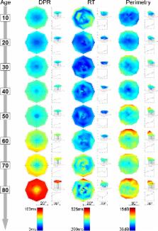
          Temporal performance parameters vary across the visual field. Their topographical distributions relative to each other and relative to basic visual performance measures and their relative change over the life span are unknown. Our goal was to characterize the topography and age-related change of temporal performance. We acquired visual field maps in 95 healthy participants (age: 10–90 years): perimetric thresholds, double-pulse resolution (DPR), reaction times (RTs), and letter contrast thresholds. DPR and perimetric thresholds increased with eccentricity and age; the periphery showed a more pronounced age-related increase than the center. RT increased only slightly and uniformly with eccentricity. It remained almost constant up to the age of 60, a marked change occurring only above 80. Overall, age was a poor predictor of functionality. Performance decline could be explained only in part by the aging of the retina and optic media. In Part II, we therefore examine higher visual and cognitive functions.

          Electronic supplementary material

          The online version of this article (doi:10.3758/s13414-012-0278-z) contains supplementary material, which is available to authorized users.

          Related collections

          Most cited references69

          • Record: found
          • Abstract: found
          • Article: not found

          The inner experience of time.

          The striking diversity of psychological and neurophysiological models of 'time perception' characterizes the debate on how and where in the brain time is processed. In this review, the most prominent models of time perception will be critically discussed. Some of the variation across the proposed models will be explained, namely (i) different processes and regions of the brain are involved depending on the length of the processed time interval, and (ii) different cognitive processes may be involved that are not necessarily part of a core timekeeping system but, nevertheless, influence the experience of time. These cognitive processes are distributed over the brain and are difficult to discern from timing mechanisms. Recent developments in the research on emotional influences on time perception, which succeed decades of studies on the cognition of temporal processing, will be highlighted. Empirical findings on the relationship between affect and time, together with recent conceptualizations of self- and body processes, are integrated by viewing time perception as entailing emotional and interoceptive (within the body) states. To date, specific neurophysiological mechanisms that would account for the representation of human time have not been identified. It will be argued that neural processes in the insular cortex that are related to body signals and feeling states might constitute such a neurophysiological mechanism for the encoding of duration.
            Bookmark
            • Record: found
            • Abstract: found
            • Article: not found

            Neuropsychology of timing and time perception.

            Interval timing in the range of milliseconds to minutes is affected in a variety of neurological and psychiatric populations involving disruption of the frontal cortex, hippocampus, basal ganglia, and cerebellum. Our understanding of these distortions in timing and time perception are aided by the analysis of the sources of variance attributable to clock, memory, decision, and motor-control processes. The conclusion is that the representation of time depends on the integration of multiple neural systems that can be fruitfully studied in selected patient populations.
              Bookmark
              • Record: found
              • Abstract: found
              • Article: not found

              Neural bases of visual deficits during aging.

              Visual abilities decline during normal (non-pathological) aging. Many of these visual declines cannot be attributed to optical changes and must therefore be due to changes in the retina or central visual pathways. These include declines in visual acuity and spatial contrast sensitivity (especially under low luminance levels), suprathreshold contrast vision and contrast gain, temporal-frequency contrast sensitivity and resolution, spatial-temporal interactions, hyperacuity, binocular processing, and sensitivity to motion. Certain aspects of these vision deficits and comparisons with neurophysiological and lesion-behavior studies in monkeys suggest hypotheses about the nature and location (e.g. magnocellular vs parvocellular pathways, specific visual structures, and so on) of the neural deficits. Despite the well-documented psychophysical deficits, available anatomical studies in humans and monkeys suggest that aging has only relatively minor effects on the retino-geniculo-striate pathway. Retinal photoreceptor losses are relatively restricted to rods, and there is compensation among the remaining rods for those that are lost. Although some retinal ganglion cells appear to be lost, the loss is small relative to individual-to-individual variability. In addition, there appear to be no massive cell losses in the LGN or striate cortex. Physiological results in the monkey LGN suggest that the functional properties of LGN neurons, and therefore their retinal inputs, are not significantly affected by aging. Retinal pattern-evoked ERG studies in humans likewise suggest that the physiological properties of the retina are little affected by aging. Comparisons between pattern-evoked ERG and cortical evoked potentials in the same individuals suggest that some neural change occurs between the retina and striate cortex, but the location and nature of this change is not known. Thus, we are far from being able to answer the question, What are the neural bases of visual deficits during aging? There are several possible reasons for this: (1) The neurobiological methods that have been brought to bear on the question have been fairly limited. (2) Investigations of neural changes may not have been guided sufficiently by what is known about the psychophysical changes that occur with aging. (3) Existing studies may not have examined the correct locations in the visual system. (4) There is large individual-to-individual variability in the effects of aging and, with the small samples of individuals that typically are available in neural studies of aging, the variability could obscure detection of aging-related changes. Suggestions are offered for ways in which future research can solve these problems.(ABSTRACT TRUNCATED AT 400 WORDS)
                Bookmark

                Author and article information

                Contributors
                +49-4221-9160109 , dapoggel@gmail.com
                strasburger@uni-muenchen.de
                Journal
                Atten Percept Psychophys
                Atten Percept Psychophys
                Attention, Perception & Psychophysics
                Springer-Verlag (New York )
                1943-3921
                1943-393X
                7 April 2012
                7 April 2012
                2012
                : 74
                : 6
                : 1114-1132
                Affiliations
                [1 ]GRID grid.5252.0, ISNI 000000041936973X, Generation Research Program (GRP), , Ludwig-Maximilian University Munich, Human Science Center, ; Bad Tölz, Germany
                [2 ]GRID grid.7450.6, ISNI 0000000123644210, Department of Medical Psychology and Medical Sociology, , Georg-August University Göttingen, ; Göttingen, Germany
                [3 ]GRID grid.5252.0, ISNI 000000041936973X, IuK, , Ludwig-Maximilian University Munich, ; Munich, Germany
                [4 ]Hanse-Wissenschaftskolleg Institute of Advanced Study, Lehmkuhlenbusch 4, 27753 Delmenhorst, Germany
                Article
                278
                10.3758/s13414-012-0278-z
                5486645
                22484795
                47a5638e-44a3-4e1c-9177-837e0d248dbb
                © The Author(s) 2012
                History
                Categories
                Article
                Custom metadata
                © Psychonomic Society, Inc. 2012

                Clinical Psychology & Psychiatry
                temporal processing,visual field maps,double-pulse resolution,reaction time,perimetric thresholds,letter contrast sensitivity,visual system,aging,relative defect

                Comments

                Comment on this article