19
views
0
recommends
+1 Recommend
0 collections
    0
    shares
      • Record: found
      • Abstract: found
      • Article: found
      Is Open Access

      Post-Natal Dynamic Changes in Circulating Follicle-Stimulating Hormone, Luteinizing Hormone, Immunoreactive Inhibin, Progesterone, Testosterone and Estradiol-17β in Thoroughbred Colts until 6 Months of Age

      research-article

      Read this article at

      Bookmark
          There is no author summary for this article yet. Authors can add summaries to their articles on ScienceOpen to make them more accessible to a non-specialist audience.

          Abstract

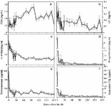
          The aim of present study was to clarify the post-natal profile of follicle-stimulating hormone (FSH), luteinizing hormone (LH), immunoreactive (ir)-inhibin, progesterone, testosterone, and estradiol-17β, and their relationships in Thoroughbred colts. Six hundred and thirty-six colts were used for the study. Single plasma samples from each animal were harvested from the blood drawn through jugular venipuncture. The subjects were born with high amounts of progesterone, testosterone, and estradiol-17β, all of which dropped significantly and remained at lower levels till the end of 6 months. FSH decreased transiently after birth until day 12 and then gradually increased to peak at day 100 which then maintained in lesser levels towards the end of the studied period. LH was highest during birth which decreased until day 26 and then increased slowly to sub-birth levels up to day 90. Animals were born with high amounts of ir-inhibin. It dropped slowly and halved by day 20 and then decreased towards rest of the studied period. The increase in FSH is negatively correlated with the declining ir-inhibin levels. The early increase in FSH can be the indication of early post-natal maturation of the hypothalamic pituitary testicular axis that ultimately might be responsible for priming the testes for future development.

          Related collections

          Most cited references18

          • Record: found
          • Abstract: found
          • Article: not found

          Role of Sertoli cell number and function on regulation of spermatogenesis.

          Testicular function is under the control of expression and repression of several genes and gene products, and many of these works through Sertoli cells. The capability of Sertoli cells to regulate spermatogenesis is dependent on Sertoli cell functions and Sertoli cell number. Sertoli cell number has long been thought to be stable in adults with no proliferation of Sertoli cells once adult numbers have been reached. However, adult horses do not have stable Sertoli cell numbers, and new studies indicate that adult Sertoli cells can be made to re-enter mitotic phase under certain experimental conditions. This review discusses roles of Sertoli cells in regulation of spermatogenesis and methods for estimating the number of Sertoli cells, in a testis, that overcome the problems (assumptions) associated with the indented, pear-shaped of Sertoli cell nuclei which make it difficult to estimate the volume of individual nuclei. Using several approaches to overcome the problems associated with any one method, the horse is identified as a species in which Sertoli cell number is not fixed, but it fluctuates with season. In addition to Sertoli cell numbers, the functions of Sertoli cells that are very important in signaling and controlling spermatogenesis are discussed. Recent studies have shown that "post-mitotic terminally differentiated Sertoli cells" from adult animals could, under certain conditions, re-enter the cell division cycle. Can seasonal influences be a natural set of conditions to induce the Sertoli cells of the horse testis to seasonally re-enter the cell division cycle and explain the seasonal differences in Sertoli cell number as summarized in this review? Alternatively, can seasonal differences in Sertoli cell number reflect, in the horse to a greater extent, but in adults of most species, the presence of some mitotic-capable Sertoli cells in adults? In any case, both Sertoli cell number and function are important in regulation of spermatogenesis.
            Bookmark
            • Record: found
            • Abstract: found
            • Article: not found

            Radioimmunoassay of inhibin in various mammals.

            A sensitive radioimmunoassay (RIA) for the determination of inhibin in peripheral plasma and tissue homogenates of different species has been developed using antisera to partially purified bovine follicular fluid (bFF) inhibin and 125I-labelled bFF 32 kDa inhibin. Antisera were produced by immunization of rabbits with partially purified bFF inhibin prepared by immunoaffinity chromatography. Increasing doses of a high titre antiserum could neutralize the suppressing effect of bFF, porcine follicular fluid and rat ovarian homogenate on FSH secretion from rat anterior pituitary cells in culture. Sensitivity of the assay was 3.1 ng International Research Standard of porcine inhibin per tube. Parallel inhibition curves were obtained for inhibin preparations from female and male animals of ten species, i.e. cattle, goats, sheep, cats, dogs, monkeys, pigs, horses, rats and man. Inhibin subunits and related proteins cross-reacted minimally with the antiserum used in the study. Plasma concentrations of inhibin in adult male and female rats were measured by the RIA before and at various times after gonadectomy. Inhibin levels in peripheral plasma before gonadectomy were significantly higher in adult female than in adult male rats. Inhibin levels decreased abruptly after gonadectomy in both sexes and they correlated negatively with plasma concentrations of FSH. This inhibin RIA will facilitate studies of the physiology of inhibin in various species of animals.
              Bookmark
              • Record: found
              • Abstract: found
              • Article: not found

              Antral follicle growth and endocrine changes in prepubertal cattle, sheep and goats.

              In the growing heifer calve, there is an early post-natal, gonadotrophin driven increase in ovarian antral follicle growth. The endocrine regulation of and reason for this initial stimulation of ovarian follicular development are not fully understood. This initial endocrine activity appears to be later held in check by negative feedback suppression mechanisms until the heifer is of a sufficient body size to initiate oestrous cycles and to reproduce. There is increasing evidence from recent ultrasonographic studies, performed in the same groups of prepubertal heifer calves, that the development of ovarian antral follicles and tubular genitalia occur in parallel. There appear to be two distinct periods of enhanced development of the reproductive organs, from 2 to 14 weeks of age and again from 34 to 60 weeks of age, or just prior to puberty. First ovulation in heifers is preceded by a gradual increase in pulsed LH secretion, which results in enhanced antral follicle development and oestrogen production. It was demonstrated that prepubertal heifers produced recurrent antral follicular waves; maximum sizes and life span of the dominant follicles of waves, as well as periodicity and FSH dependency of wave emergence were similar to those in adult cattle. In does, no Graafian follicles are seen at birth and total follicle numbers increase to 2 months of age, and then decline to 5 months of age. In ewe lambs, studies using transrectal ovarian ultrasonography showed that antral follicle recruitment and growth increased after the first 2 months of age and just before puberty. This bi-phasic pattern of changes in ovarian follicle recruitment and growth is strikingly similar to that in heifer calves, but it contrasts with earlier post-mortem examinations of ovaries in ewe lambs. Unlike in cattle and adult ewes, the rhythmic pattern of follicular wave emergence was not established in pre- and peripubertal ewe lambs. The early increase in antral follicle numbers and size in ewe lambs may be, at least in part, due to changes in FSH release and potency, and enhanced follicle production prior to first ovulation is probably caused by an increase in the frequency of LH pulses.
                Bookmark

                Author and article information

                Journal
                J Equine Sci
                J Equine Sci
                JES
                Journal of equine science
                The Japanese Society of Equine Science
                1340-3516
                1347-7501
                26 April 2011
                2011
                : 22
                : 1
                : 9-15
                Affiliations
                [1 ]Department of Basic Veterinary Science, United Graduate School of Veterinary Sciences, Gifu University, Gifu 501-1193, Japan
                [2 ]Laboratory of Veterinary Physiology, Department of Veterinary Medicine, Faculty of Agriculture, Tokyo University of Agriculture and Technology, Tokyo 183-8509, Japan
                [3 ]Shadai Corporation, Hokkaido 059-1432, Japan
                [4 ]Hidaka Training Research Center, Japan Racing Association, Hokkaido 057-0171, Japan
                [5 ]Department of Veterinary Pathology, Rakuno Gakuen University, Hokkaido 069-850, Japan
                Author notes
                [* ] Corresponding author. e-mail: taya@ 123456cc.tuat.ac.jp

                P.DHAKAL and N.TSUNODA contributed equally to this study.

                Article
                1015
                10.1294/jes.22.9
                4013995
                4810d9aa-3acb-4bac-8fb1-d08724700fbb
                2011 The Japanese Society of Equine Science

                This is an open-access article distributed under the terms of the Creative Commons Attribution Non-Commercial No Derivatives (by-nc-nd) License.

                History
                : 20 December 2010
                Categories
                Original Article

                fsh,ir-inhibin,post-natal,steroid hormones,thoroughbred colt

                Comments

                Comment on this article