16
views
0
recommends
+1 Recommend
0 collections
    0
    shares
      • Record: found
      • Abstract: found
      • Article: not found

      Evidence against dopamine D1/D2 receptor heteromers

      research-article

      Read this article at

      ScienceOpenPublisherPMC
      Bookmark
          There is no author summary for this article yet. Authors can add summaries to their articles on ScienceOpen to make them more accessible to a non-specialist audience.

          Abstract

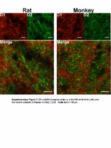
          Hetero-oligomers of G-protein-coupled receptors have become the subject of intense investigation because their purported potential to manifest signaling and pharmacological properties that differ from the component receptors makes them highly attractive for the development of more selective pharmacological treatments. In particular, dopamine D1 and D2 receptors have been proposed to form hetero-oligomers that couple to G αq proteins, and SKF83959 has been proposed to act as a biased agonist that selectively engages these receptor complexes to activate G αq and thus phospholipase C. D1/D2 heteromers have been proposed as relevant to the pathophysiology and treatment of depression and schizophrenia. We used in vitro bioluminescence resonance energy transfer (BRET), ex vivo analyses of receptor localization and proximity in brain slices, and behavioral assays in mice to characterize signaling from these putative dimers/oligomers. We were unable to detect G αq or G α11 protein coupling to homomers or heteromers of D1 or D2 receptors using a variety of biosensors. SKF83959-induced locomotor and grooming behaviors were eliminated in D 1 receptor knockout mice, verifying a key role for D1-like receptor activation. In contrast, SKF83959-induced motor responses were intact in D2 receptor and G αq knockout mice, as well as in knock-in mice expressing a mutant Ala 286-CaMKIIα, that cannot autophosphorylate to become active. Moreover, we found that in the shell of the nucleus accumbens, even in neurons in which D1 and D2 receptor promoters are both active, the receptor proteins are segregated and do not form complexes. These data are not compatible with SKF83959 signaling through G αq or through a D1–D2 heteromer and challenge the existence of such a signaling complex in the adult animals that we used for our studies.

          Related collections

          Most cited references68

          • Record: found
          • Abstract: found
          • Article: not found

          A gene expression atlas of the central nervous system based on bacterial artificial chromosomes.

          The mammalian central nervous system (CNS) contains a remarkable array of neural cells, each with a complex pattern of connections that together generate perceptions and higher brain functions. Here we describe a large-scale screen to create an atlas of CNS gene expression at the cellular level, and to provide a library of verified bacterial artificial chromosome (BAC) vectors and transgenic mouse lines that offer experimental access to CNS regions, cell classes and pathways. We illustrate the use of this atlas to derive novel insights into gene function in neural cells, and into principal steps of CNS development. The atlas, library of BAC vectors and BAC transgenic mice generated in this screen provide a rich resource that allows a broad array of investigations not previously available to the neuroscience community.
            Bookmark
            • Record: found
            • Abstract: found
            • Article: not found

            Autophosphorylation at Thr286 of the alpha calcium-calmodulin kinase II in LTP and learning.

            The calcium-calmodulin-dependent kinase II (CaMKII) is required for hippocampal long-term potentiation (LTP) and spatial learning. In addition to its calcium-calmodulin (CaM)-dependent activity, CaMKII can undergo autophosphorylation, resulting in CaM-independent activity. A point mutation was introduced into the alphaCaMKII gene that blocked the autophosphorylation of threonine at position 286 (Thr286) of this kinase without affecting its CaM-dependent activity. The mutant mice had no N-methyl-D-aspartate receptor-dependent LTP in the hippocampal CA1 area and showed no spatial learning in the Morris water maze. Thus, the autophosphorylation of alphaCaMKII at Thr286 appears to be required for LTP and learning.
              Bookmark
              • Record: found
              • Abstract: found
              • Article: not found

              Opposing patterns of signaling activation in dopamine D1 and D2 receptor-expressing striatal neurons in response to cocaine and haloperidol.

              Psychostimulants and other drugs of abuse activate extracellular signal-regulated kinase (ERK) in the striatum, through combined stimulation of dopamine D(1) receptors (D1Rs) and glutamate NMDA receptors. Antipsychotic drugs activate similar signaling proteins in the striatum by blocking dopamine D(2) receptors (D2Rs). However, the neurons in which these pathways are activated by psychotropic drugs are not precisely identified. We used transgenic mice, in which enhanced green fluorescent protein (EGFP) expression was driven by D1R promoter (drd1a-EGFP) or D2R promoter (drd2-EGFP). We confirmed the expression of drd1a-EGFP in striatonigral and drd2-EGFP in striatopallidal neurons. Drd2-EGFP was also expressed in cholinergic interneurons, whereas no expression of either promoter was detected in GABAergic interneurons. Acute cocaine treatment increased phosphorylation of ERK and its direct or indirect nuclear targets, mitogen- and stress-activated kinase-1 (MSK1) and histone H3, exclusively in D1R-expressing output neurons in the dorsal striatum and nucleus accumbens. Cocaine-induced expression of c-Fos and Zif268 predominated in D1R-expressing neurons but was also observed in D2R-expressing neurons. One week after repeated cocaine administration, cocaine-induced signaling responses were decreased, with the exception of enhanced ERK phosphorylation in dorsal striatum. The responses remained confined to D1R neurons. In contrast, acute haloperidol injection activated phosphorylation of ERK, MSK1, and H3 only in D2R neurons and induced c-fos and zif268 predominantly in these neurons. Our results demonstrate that cocaine and haloperidol specifically activate signaling pathways in two completely segregated populations of striatal output neurons, providing direct evidence for the selective mechanisms by which these drugs exert their long-term effects.
                Bookmark

                Author and article information

                Journal
                9607835
                20545
                Mol Psychiatry
                Mol. Psychiatry
                Molecular psychiatry
                1359-4184
                1476-5578
                30 October 2014
                06 January 2015
                November 2015
                01 May 2016
                : 20
                : 11
                : 1373-1385
                Affiliations
                [1 ]Neuroscience Graduate Program, Vanderbilt University School of Medicine, Nashville, TN 37232, USA
                [2 ]Departments of Psychiatry and Pharmacology, College of Physicians and Surgeons, Columbia University, New York, New York, USA
                [3 ]Nutrition and Integrative Neurobiology, INRA UMR 1286; University of Bordeaux, F-33076, Bordeaux, France
                [4 ]Center for Neuroscience. Columbia University, Kolb Research Building, New York, NY10032, USA
                [5 ]Molecular Neuropharmacology Section, National Institute of Neurological Disorders and Stroke, National Institutes of Health, Bethesda, MD, USA
                [6 ]Department of Psychiatry, McLean Hospital, Harvard Medical School, Belmont, MA, USA
                [7 ]Department of Pharmacology, Vanderbilt University School of Medicine, Nashville, TN 37232, USA
                [8 ]Department of Molecular Physiology and Biophysics, Vanderbilt University School of Medicine, Nashville, TN 37232, USA
                [9 ]Vanderbilt Kennedy Center and Vanderbilt Brain Institute, Vanderbilt University School of Medicine, Nashville, TN 37232, USA
                [10 ]Division of Molecular Therapeutics, New York State Psychiatric Institute, New York, New York, USA
                Author notes
                Correspondence to either: Gregg D. Stanwood, Ph.D., Department of Pharmacology, 8405 MRBIV, 2213 Garland Ave, Nashville, TN 37232-6600, Tel: (615) 936-3861, gregg.stanwood@ 123456vanderbilt.edu , Jonathan A. Javitch, M.D./Ph.D., Columbia University/New York State Psychiatric Institute, 1051 Riverside Drive, Unit 19, New York, NY 10032, Tel: (646) 774-8600, jaj2@ 123456columbia.edu
                [11]

                Current address: National Institute on Drug Abuse, National Institutes of Health, Baltimore, MD 21224

                [*]

                these authors contributed equally to this work

                Article
                NIHMS638379
                10.1038/mp.2014.166
                4492915
                25560761
                481ef65f-d086-46f9-8d6e-e8af86e384cf

                Users may view, print, copy, and download text and data-mine the content in such documents, for the purposes of academic research, subject always to the full Conditions of use: http://www.nature.com/authors/editorial_policies/license.html#terms

                History
                Categories
                Article

                Molecular medicine
                dopamine,gαq,biased agonism,hetero-oligomer,d1,d2,d5,bret,proximity ligation assay,striatum
                Molecular medicine
                dopamine, gαq, biased agonism, hetero-oligomer, d1, d2, d5, bret, proximity ligation assay, striatum

                Comments

                Comment on this article