64
views
0
recommends
+1 Recommend
0 collections
    0
    shares
      • Record: found
      • Abstract: found
      • Article: found
      Is Open Access

      An activator of mTOR inhibits oxLDL-induced autophagy and apoptosis in vascular endothelial cells and restricts atherosclerosis in apolipoprotein E -/- mice

      research-article

      Read this article at

      Bookmark
          There is no author summary for this article yet. Authors can add summaries to their articles on ScienceOpen to make them more accessible to a non-specialist audience.

          Abstract

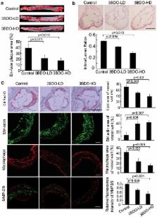
          Oxidized low-density lipoprotein (oxLDL) inhibits mammalian target of rapamycin (mTOR) and induces autophagy and apoptosis in vascular endothelial cells (VECs) that play very critical roles for the cardiovascular homostasis. We recently defined 3-benzyl-5-((2-nitrophenoxy) methyl)-dihydrofuran-2(3H)-one (3BDO) as a new activator of mTOR. Therefore, we hypothesized that 3BDO had a protective role in VECs and thus stabilized atherosclerotic lesions in apolipoprotein E -/- (apoE -/-) mice. Our results showed that oxLDL inhibited the activity of mTOR and increased the protein level of autophagy-related 13 (ATG13) and its dephosphorylation, thus inducing autophagy in human umbilical vein endothelial cells (HUVECs). All of these effects were strongly inhibited by 3BDO. In vivo experiments confirmed that 3BDO activated mTOR and decreased the protein level of ATG13 in the plaque endothelium of apoE -/- mice. Importantly, 3BDO did not affect the activity of mTOR and autophagy in macrophage cell line RAW246.7 and vascular smooth muscle cells of apoE -/- mice, but suppressed plaque endothelial cell death and restricted atherosclerosis development in the mice. 3BDO protected VECs by activating mTOR and thus stabilized atherosclerotic lesions in apoE -/- mice.

          Related collections

          Most cited references33

          • Record: found
          • Abstract: found
          • Article: not found

          Autophagy in cell death: an innocent convict?

          The visualization of autophagosomes in dying cells has led to the belief that autophagy is a nonapoptotic form of programmed cell death. This concept has now been evaluated using cells and organisms deficient in autophagy genes. Most evidence indicates that, at least in cells with intact apoptotic machinery, autophagy is primarily a pro-survival rather than a pro-death mechanism. This review summarizes the evidence linking autophagy to cell survival and cell death, the complex interplay between autophagy and apoptosis pathways, and the role of autophagy-dependent survival and death pathways in clinical diseases.
            Bookmark
            • Record: found
            • Abstract: found
            • Article: not found

            A novel, human Atg13 binding protein, Atg101, interacts with ULK1 and is essential for macroautophagy.

            Macroautophagy is an intracellular, vesicle-mediated mechanism for the sequestration and ultimate lysosomal degradation of cytoplasmic proteins, organelles and macromolecules. The macroautophagy process and many of the autophagy-specific (Atg) proteins are remarkably well conserved in higher eukaryotes. In yeast, the Atg1 kinase complex includes Atg1, Atg13, Atg17, and at least four other interacting proteins, some of which are phosphorylated in a TOR-dependent manner, placing the Atg1 signaling complex downstream of a major nutrient-sensing pathway. Atg1 orthologs, including mammalian unc-51-like kinase 1 (ULK1), have been identified in higher eukaryotes and have been functionally linked to autophagy. This suggests that other components of the Atg1 complex exist in higher eukaryotes. Recently, a putative human Atg13 ortholog, FLJ20698, was identified by gapped-BLAST analysis. We show here that FLJ20698 (Atg13) is a ULK1-interacting phosphoprotein that is essential for macroautophagy. Furthermore, we identify a novel, human Atg13-interacting protein, FLJ11773, which we have termed Atg101. Atg101 is essential for autophagy and interacts with ULK1 in an Atg13-dependent manner. Additionally, we present evidence that intracellular localization of the ULK1 complex is regulated by nutrient conditions. Finally, we demonstrate that Atg101 stabilizes the expression of Atg13 in the cell, suggesting that Atg101 contributes to Atg13 function by protecting Atg13 from proteasomal degradation. Therefore, the identification of the novel protein, Atg101, and the validation of Atg13 and Atg101 as ULK1-interacting proteins, suggests an Atg1 complex is involved in the induction of macroautophagy in mammalian cells.
              Bookmark
              • Record: found
              • Abstract: found
              • Article: not found

              Hypoxia enhances vascular cell proliferation and angiogenesis in vitro via rapamycin (mTOR)-dependent signaling.

              Angiogenesis and vascular cell proliferation are pivotal in physiological and pathological processes including atherogenesis, restenosis, wound healing, and cancer development. Here we show that mammalian target of rapamycin (mTOR) signaling plays a key role in hypoxia-triggered smooth muscle and endothelial proliferation and angiogenesis in vitro. Hypoxia significantly increased DNA synthesis and proliferative responses to platelet-derived growth factor (PDGF) and fibroblast growth factor (FGF) in rat and human smooth muscle and endothelial cells. In an in vitro 3-dimensional model of angiogenesis, hypoxia increased PDGF- and FGF-stimulated sprout formation from rat and mouse aortas. Hypoxia did not modulate PDGF receptor mRNA, protein, or phosphorylation. PI3K activity was essential for cell proliferation under normoxic and hypoxic conditions. Activities of PI3K-downstream target PKB under hypoxia and normoxia were comparable. However, mTOR inhibition by rapamycin specifically abrogated hypoxia-mediated amplification of proliferation and angiogenesis, but was without effect on proliferation under normoxia. Accordingly, hypoxia-mediated amplification of proliferation was further augmented in mTOR-overexpressing endothelial cells. Thus, signaling via mTOR may represent a novel mechanism whereby hypoxia augments mitogen-stimulated vascular cell proliferation and angiogenesis.
                Bookmark

                Author and article information

                Journal
                Sci Rep
                Sci Rep
                Scientific Reports
                Nature Publishing Group
                2045-2322
                01 July 2014
                2014
                : 4
                : 5519
                Affiliations
                [1 ]Shandong Provincial Key Laboratory of Animal Cells and Developmental Biology, School of Life Science, Shandong University , Jinan 250100, China
                [2 ]School of Biological Science and Technology, University of Jinan , Jinan 250022, China
                [3 ]Institute of Organic Chemistry, School of Chemistry and Chemical Engineering, Shandong University , Jinan 250100, China
                [4 ]The Key Laboratory of Cardiovascular Remodeling and Function Research, Chinese Ministry of Education and Chinese Ministry of Health, Shandong University Qilu Hospital , Jinan, 250012, China
                [5 ]These authors contributed equally to this work.
                Author notes
                Article
                srep05519
                10.1038/srep05519
                4076681
                24980430
                4975310b-c516-41a9-84a9-e72877848979
                Copyright © 2014, Macmillan Publishers Limited. All rights reserved

                This work is licensed under a Creative Commons Attribution-NonCommercial-NoDerivs 4.0 International License. The images or other third party material in this article are included in the article's Creative Commons license, unless indicated otherwise in the credit line; if the material is not included under the Creative Commons license, users will need to obtain permission from the license holder in order to reproduce the material. To view a copy of this license, visit http://creativecommons.org/licenses/by-nc-nd/4.0/

                History
                : 31 March 2014
                : 13 June 2014
                Categories
                Article

                Uncategorized
                Uncategorized

                Comments

                Comment on this article