1
views
0
recommends
+1 Recommend
0 collections
    0
    shares
      • Record: found
      • Abstract: found
      • Article: found
      Is Open Access

      Efficacy of a Second Brain Biopsy for Intracranial Lesions after Initial Negativity

      research-article

      Read this article at

      Bookmark
          There is no author summary for this article yet. Authors can add summaries to their articles on ScienceOpen to make them more accessible to a non-specialist audience.

          Abstract

          Background and Purpose

          The rationale for performing a second brain biopsy after initial negativity is not well evaluated in the literature. This study was designed to 1) assess the efficacy of a second brain biopsy when the first biopsy was nondiagnostic, 2) identify possible factors associated with an increased diagnostic rate in the second biopsy, and 3) analyze additional morbidity induced by the second biopsy.

          Methods

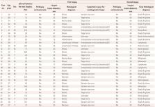
          We performed a retrospective cohort study from 2009 to 2019, during which 1,919 patients underwent a brain biopsy, including 30 who were biopsied twice (1.6%). The specific histological diagnosis rate, diagnosis-associated factors, and complication rate were assessed for the 30 twice-biopsied patients.

          Results

          The second biopsy allowed a specific histological diagnosis in 86.7% of the patients who had initially undergone a nondiagnostic brain biopsy [odds ratio (OR)=7.5, 95% confidence interval (CI)=3.0–18.7, p<0.001]. The multivariate analysis showed that only prebiopsy corticosteroid administration (OR=2.6, 95% CI=1.1–6.0, p=0.01) was an important factor in predicting a nondiagnostic biopsy. None of the patients developed a symptomatic complication after the first biopsy, while two (6.0%) patients experienced a transient complication after the second biopsy ( p=0.49).

          Conclusions

          Performing a second brain biopsy in patients who have an initial nondiagnostic biopsy is effective in most cases. We advocate that a second biopsy be systematically considered in the diagnosis algorithm of these patients after it has been verified that molecular testing cannot help to obtain a diagnosis. Corticosteroid administration can lead to nondiagnostic biopsies and should be avoided when possible during the prebiopsy period.

          Related collections

          Most cited references35

          • Record: found
          • Abstract: found
          • Article: not found

          Clinical Metagenomic Sequencing for Diagnosis of Meningitis and Encephalitis

          BACKGROUND Metagenomic next-generation sequencing (NGS) of cerebrospinal fluid (CSF) has the potential to identify a broad range of pathogens in a single test. METHODS In a 1-year, multicenter, prospective study, we investigated the usefulness of metagenomic NGS of CSF for the diagnosis of infectious meningitis and encephalitis in hospitalized patients. All positive tests for pathogens on metagenomic NGS were confirmed by orthogonal laboratory testing. Physician feedback was elicited by teleconferences with a clinical microbial sequencing board and by surveys. Clinical effect was evaluated by retrospective chart review. RESULTS We enrolled 204 pediatric and adult patients at eight hospitals. Patients were severely ill: 48.5% had been admitted to the intensive care unit, and the 30-day mortality among all study patients was 11.3%. A total of 58 infections of the nervous system were diagnosed in 57 patients (27.9%). Among these 58 infections, metagenomic NGS identified 13 (22%) that were not identified by clinical testing at the source hospital. Among the remaining 45 infections (78%), metagenomic NGS made concurrent diagnoses in 19. Of the 26 infections not identified by metagenomic NGS, 11 were diagnosed by serologic testing only, 7 were diagnosed from tissue samples other than CSF, and 8 were negative on metagenomic NGS owing to low titers of pathogens in CSF. A total of 8 of 13 diagnoses made solely by metagenomic NGS had a likely clinical effect, with 7 of 13 guiding treatment. CONCLUSIONS Routine microbiologic testing is often insufficient to detect all neuroinvasive pathogens. In this study, metagenomic NGS of CSF obtained from patients with meningitis or encephalitis improved diagnosis of neurologic infections and provided actionable information in some cases. (Funded by the National Institutes of Health and others; PDAID ClinicalTrials.gov number, [Related object:] .)
            Bookmark
            • Record: found
            • Abstract: found
            • Article: not found

            Frequency of TERT promoter mutations in human cancers.

            Reactivation of telomerase has been implicated in human tumorigenesis, but the underlying mechanisms remain poorly understood. Here we report the presence of recurrent somatic mutations in the TERT promoter in cancers of the central nervous system (43%), bladder (59%), thyroid (follicular cell-derived, 10%) and skin (melanoma, 29%). In thyroid cancers, the presence of TERT promoter mutations (when occurring together with BRAF mutations) is significantly associated with higher TERT mRNA expression, and in glioblastoma we find a trend for increased telomerase expression in cases harbouring TERT promoter mutations. Both in thyroid cancers and glioblastoma, TERT promoter mutations are significantly associated with older age of the patients. Our results show that TERT promoter mutations are relatively frequent in specific types of human cancers, where they lead to enhanced expression of telomerase.
              Bookmark
              • Record: found
              • Abstract: found
              • Article: not found

              Corticosteroids and the brain

              The brain is continuously exposed to varying levels of adrenal corticosteroid hormones such as corticosterone in rodents and cortisol in humans. Natural fluctuations occur due to ultradian and circadian variations or are caused by exposure to stressful situations. Brain cells express two types of corticosteroid receptors, i.e. mineralocorticoid and glucocorticoid receptors, which differ in distribution and affinity. These receptors can mediate both rapid non-genomic and slow gene-mediated neuronal actions. As a consequence of these factors, natural (e.g. stress-induced) shifts in corticosteroid level are associated with a complex mosaic of time- and region-dependent changes in neuronal activity. A series of experiments in humans and rodents have revealed that these time- and region-dependent cellular characteristics are also reflected in distinct cognitive patterns after stress. Thus, directly after a peak of corticosteroids, attention and vigilance are increased, and areas involved in emotional responses and simple behavioral strategies show enhanced activity. In the aftermath of stress, areas involved in higher cognitive functions become activated allowing individuals to link stressful events to the specific context and to store information for future use. Both phases of the brain’s response to stress are important to face a continuously changing environment, promoting adaptation at the short as well as long term. We argue that a balanced response during the two phases is essential for resilience. This balance may become compromised after repeated stress exposure, particularly in genetically vulnerable individuals and aggravate disease manifestation. This not only applies to psychiatric disorders but also to neurological diseases such as epilepsy.
                Bookmark

                Author and article information

                Journal
                J Clin Neurol
                J Clin Neurol
                JCN
                Journal of Clinical Neurology (Seoul, Korea)
                Korean Neurological Association
                1738-6586
                2005-5013
                October 2020
                25 September 2020
                : 16
                : 4
                : 659-667
                Affiliations
                [a ]Department of Neurosurgery, La Pitié-Salpêtrière-Charles Foix University Hospital, Assistance Publique-Hôpitaux de Paris, Paris, France.
                [b ]Department of Neuropathology, La Pitié-Salpêtrière-Charles Foix University Hospital, Assistance Publique-Hôpitaux de Paris, Paris, France.
                [c ]Sorbonne University, Paris, France.
                [d ]Paris Brain Institute, Paris, France.
                [e ]Department of Neuro-Oncology, La Pitié-Salpêtrière-Charles Foix University Hospital, Assistance Publique-Hôpitaux de Paris, Paris, France.
                Author notes
                Correspondence: Bertrand Mathon, MD, MSc. Department of Neurosurgery, La Pitié-Salpêtrière University Hospital, 47-83, boulevard de l'Hôpital, 75651 Paris Cedex 13, France. Tel +33-1-84-82-73-63, Fax +33-1-42-16-34-08, bertrand.mathon@ 123456aphp.fr
                Author information
                https://orcid.org/0000-0001-7760-9226
                https://orcid.org/0000-0003-3659-291X
                https://orcid.org/0000-0001-6564-6388
                https://orcid.org/0000-0001-7312-6358
                https://orcid.org/0000-0002-5910-7799
                https://orcid.org/0000-0002-9182-5846
                Article
                10.3988/jcn.2020.16.4.659
                7542000
                33029973
                4b21e72b-a819-430e-940e-3d9483a7a6a7
                Copyright © 2020 Korean Neurological Association

                This is an Open Access article distributed under the terms of the Creative Commons Attribution Non-Commercial License ( http://creativecommons.org/licenses/by-nc/4.0/) which permits unrestricted non-commercial use, distribution, and reproduction in any medium, provided the original work is properly cited.

                History
                : 19 May 2020
                : 21 July 2020
                : 21 July 2020
                Categories
                Original Article

                Neurology
                brain tumor,corticosteroids,diagnosis,neuropathology,neurosurgery,neoplasms
                Neurology
                brain tumor, corticosteroids, diagnosis, neuropathology, neurosurgery, neoplasms

                Comments

                Comment on this article