19
views
0
recommends
+1 Recommend
0 collections
    0
    shares
      • Record: found
      • Abstract: found
      • Article: found
      Is Open Access

      Simultaneous Bilateral Infective Endocarditis with Right Ventricular Mural Involvement

      case-report

      Read this article at

      Bookmark
          There is no author summary for this article yet. Authors can add summaries to their articles on ScienceOpen to make them more accessible to a non-specialist audience.

          Abstract

          A 47-year-old woman without a significant medical history, including no history of intravenous drug abuse, no body piercings, and no tattoos, was referred with a 6-day history of high fever and arthralgia. Blood cultures were all positive for Staphylococcus 
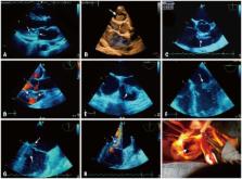
aureus Meti-S. Transthoracic echocardiography revealed: 1) an irregular thickening of the posterior mitral valve root with mobile extensions projecting toward both the left ventricle and the left atrium (Fig. 1A, Supplementary movie 1); 2) a huge mobile vegetation rising from the upper portion of the right side of the interventricular septum (Fig. 1B, Supplementary movie 2). These findings were confirmed by transesophageal echocardiography (Fig. 1C-G, Supplementary movie 3 and 4), which also disclosed a mild-to-moderate mitral regurgitation (Fig. 1H). A diagnosis of multisite infective endocarditis with right sided mural involvement was made. No point of entry was detected. The patient got quickly worse and was referred for emergency cardiac surgery.1) Intraoperative findings confirmed the presence of a bulky 3 cm-long vegetation attached to the right-sided interventricular septal endocardial surface that reached the pulmonary valve orifice (Fig. 1I, Supplementary movie 5). On the left side of the heart, the surgeon noted the presence of a large vegetation extending to the posterior free wall endocardium that was damaging the root of the mitral posterior leaflet, and destroying several chordae tendineae and the top of the posterior papillary muscle. The surgical procedure consisted of a conservative posterior mitral valve repair and, on the right side, of a single septal vegectomy. The patient was discharged home 38 days after surgery, in stable clinical condition.

          Related collections

          Most cited references1

          • Record: found
          • Abstract: found
          • Article: not found

          Surgical treatment of multivalvular endocarditis: twenty-one-year single center experience.

          Little information is available about surgical outcomes in patients with multivalvular endocarditis. The aim of this article is to review the 21-year experience with surgical treatment of patients with multivalvular endocarditis at our institution and, in particular, to determine the incidence, pathologic status, diagnosis, surgical strategies, and outcomes of patients with this disease. From January 1986 to December 2006, a total of 48 patients (40 men, 8 women), with a mean age of 42 +/- 12 years, underwent surgery for multivalvular endocarditis. Endocarditis was active in 32 patients and healed in 16. Preoperative transthoracic echocardiographic evaluation was performed in all 48 patients with addition of transesophageal echocardiography in 22 (45.8%). Intraoperative findings showed that the endocarditis involved mostly the mitral and aortic valves (40/48 patients). Triple or quadruple valve involvement was found in 1 and 2 patients, respectively. Preoperative, perioperative, and postoperative data were retrospectively analyzed and risk factors for early and late survival were determined. In only 24 (50.0%) patients was multivalvular endocarditis diagnosed by preoperative transthoracic echocardiography; 17 (77.3%) patients had multivalvular endocarditis confirmed by preoperative transesophageal echocardiography. The 30-day hospital mortality was 12.5% (n = 6). Preoperative renal failure, New York Heart Association class IV, and emergency surgery were identified as independent risk factors for hospital mortality. Overall long-term survival was 74% +/- 6% at 5 years and 62% +/- 3% at 10 years. Multivariate analysis revealed that renal failure and recurrent endocarditis were associated with increased late mortality. Ten-year freedom from recurrent endocarditis was 74% +/- 5% and 10-year freedom from reoperation was 73% +/- 6%. In our institution, multivalvular endocarditis was diagnosed by transthoracic echocardiography in only half of the patients. Intraoperative transesophageal echocardiography provided a more effective means to identify this disease. Radical resection of all infected tissues for patients with multivalvular endocarditis and additional intraoperative interventions, depending on the intraoperative pathologic condition, produced satisfactory in-hospital and long-term results, similar to those in patients with a single infected heart valve.
            Bookmark

            Author and article information

            Journal
            J Cardiovasc Ultrasound
            J Cardiovasc Ultrasound
            JCU
            Journal of Cardiovascular Ultrasound
            Korean Society of Echocardiography
            1975-4612
            2005-9655
            June 2015
            26 June 2015
            : 23
            : 2
            : 119-120
            Affiliations
            [1 ]Department of Clinical Physiology, Franco-Britannique Hospital, Levallois-Perret, France.
            [2 ]Intensive Care Unit, Franco-Britannique Hospital, Levallois-Perret, France.
            [3 ]CERIC, Ambroise-Paré Clinic, Neuilly-sur-Seine, France.
            Author notes
            Address for Correspondence: Dominique de Zuttere, Department of Clinical Physiology, Franco-Britannique Hospital, 4 rue Kléber, Levallois-Perret Cedex 92309, France. Tel: +33-1-47-59-55-01, Fax: +33-1-47-59-59-75, dominique.de.zuttere@ 123456ihfb.org
            Article
            10.4250/jcu.2015.23.2.119
            4486178
            4b2921c0-743b-4f4e-a4b9-019835de1dd4
            Copyright © 2015 Korean Society of Echocardiography

            This is an Open Access article distributed under the terms of the Creative Commons Attribution Non-Commercial License ( http://creativecommons.org/licenses/by-nc/3.0/) which permits unrestricted non-commercial use, distribution, and reproduction in any medium, provided the original work is properly cited.

            History
            : 29 November 2014
            : 28 December 2014
            : 19 May 2015
            Categories
            Images in Cardiovascular Ultrasound

            Cardiovascular Medicine
            echocardiography,endocarditis,infective endocarditis,cardiac surgery
            Cardiovascular Medicine
            echocardiography, endocarditis, infective endocarditis, cardiac surgery

            Comments

            Comment on this article