Inviting an author to review:
Find an author and click ‘Invite to review selected article’ near their name.
Search for authorsSearch for similar articles
23
views
0
recommends
+1 Recommend
0 collections
    0
    shares
      • Record: found
      • Abstract: found
      • Article: found
      Is Open Access

      Agreement of Anterior Segment Parameters Obtained From Swept-Source Fourier-Domain and Time-Domain Anterior Segment Optical Coherence Tomography

      research-article

      Read this article at

      Bookmark
          There is no author summary for this article yet. Authors can add summaries to their articles on ScienceOpen to make them more accessible to a non-specialist audience.

          Abstract

          Purpose

          To assess the interdevice agreement between swept-source Fourier-domain and time-domain anterior segment optical coherence tomography (AS-OCT).

          Methods

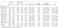
          Fifty-three eyes from 41 subjects underwent CASIA2 and Visante OCT imaging. One hundred eighty–degree axis images were measured with the built-in two-dimensional analysis software for the swept-source Fourier-domain AS-OCT (CASIA2) and a customized program for the time-domain AS-OCT (Visante OCT). In both devices, we examined the angle opening distance (AOD), trabecular iris space area (TISA), angle recess area (ARA), anterior chamber depth (ACD), anterior chamber width (ACW), and lens vault (LV). Bland-Altman plots and intraclass correlation (ICC) were performed. Orthogonal linear regression assessed any proportional bias.

          Results

          ICC showed strong correlation for LV (0.925) and ACD (0.992) and moderate agreement for ACW (0.801). ICC suggested good agreement for all angle parameters (0.771–0.878) except temporal AOD500 (0.743) and ARA750 (nasal 0.481; temporal 0.481). There was a proportional bias in nasal ARA750 (slope 2.44, 95% confidence interval [CI]: 1.95–3.18), temporal ARA750 (slope 2.57, 95% CI: 2.04–3.40), and nasal TISA500 (slope 1.30, 95% CI: 1.12–1.54). Bland-Altman plots demonstrated in all measured parameters a minimal mean difference between the two devices (−0.089 to 0.063); however, evidence of constant bias was found in nasal AOD250, nasal AOD500, nasal AOD750, nasal ARA750, temporal AOD500, temporal AOD750, temporal ARA750, and ACD. Among the parameters with constant biases, CASIA2 tends to give the larger numbers.

          Conclusions

          Both devices had generally good agreement. However, there were proportional and constant biases in most angle parameters. Thus, it is not recommended that values be used interchangeably.

          Related collections

          Most cited references27

          • Record: found
          • Abstract: found
          • Article: not found

          Detection of primary angle closure using anterior segment optical coherence tomography in Asian eyes.

          To evaluate noncontact anterior segment optical coherence technology (AS-OCT) as a qualitative method of imaging the anterior chamber angle and to determine its ability to detect primary angle closure when compared with gonioscopy in Asian subjects. Prospective observational case series. Two hundred three subjects were recruited from glaucoma clinics in Singapore with diagnoses of primary angle closure, primary open-angle glaucoma, ocular hypertension, or cataract. Both eyes (if eligible) of each patient were included in the study. Exclusion criteria were pseudophakia or previous glaucoma surgery. Images of the nasal, temporal, and inferior angles were obtained with AS-OCT in dark and then light conditions. Gonioscopic angle width was graded using the Spaeth classification for each quadrant in low lighting conditions. Angle closure was defined by AS-OCT as contact between the peripheral iris and angle wall anterior to the scleral spur and by gonioscopy as a Spaeth grade of 0 degree (posterior trabecular meshwork not visible). Comparison of the 2 methods in detecting angle closure was done by eye and by individual. Sensitivities and specificities of AS-OCT were calculated using gonioscopy as the reference standard. Complete data were available for 342 eyes of 200 patients. Of the patients, 70.9% had a clinical diagnosis of treated or untreated primary angle closure. Angle closure in > or =1 quadrants was detected by AS-OCT in 142 (71%) patients (228 [66.7%] eyes) and by gonioscopy in 99 (49.5%) patients (152 [44.4%] eyes). The inferior angle was closed more frequently than the nasal or temporal quadrants using both AS-OCT and gonioscopy. When performed under dark conditions, AS-OCT identified 98% of those subjects found to have angle closure on gonioscopy (95% confidence interval [CI], 92.2-99.6) and led to the characterization of 44.6% of those found to have open angles on gonioscopy to have angle closure as well. With gonioscopy as the reference standard, specificity of AS-OCT in the dark was 55.4% (95% CI, 45.2-65.2) for detecting individuals with angle closure. Anterior segment OCT is a rapid noncontact method of imaging angle structures. It is highly sensitive in detecting angle closure when compared with gonioscopy. More persons are found to have closed angles with AS-OCT than with gonioscopy.
            Bookmark
            • Record: found
            • Abstract: found
            • Article: not found

            Quantitative iris parameters and association with narrow angles.

            To investigate the relationship between quantitative iris parameters (iris curvature [I-Curv], iris area [I-Area], and iris thickness) and the presence of narrow angles. Cross-sectional, community-based study. We recruited 2047 subjects >50 years old without ophthalmic symptoms from a community clinic in Singapore. All subjects underwent gonioscopy and anterior segment optical coherence tomography (AS-OCT) under dark conditions. An eye was considered to have narrow angles if the posterior pigmented trabecular meshwork was not visible for >/=180 degrees on nonindentation gonioscopy with the eye in the primary position. Customized software was used on horizontal AS-OCT scans to measure I-Curv, I-Area, and iris thickness 750 mum (IT750) and 2000 mum (IT2000) from the scleral spur. The average of both temporal and nasal measured values of the right eye was used for analysis. The association between iris parameters and narrow angles on gonioscopy. Iris parameters from 1465 eyes (71.6%) were available for analysis. Of these, 315 subjects (21.5%) had narrow angles. The mean I-Curv (0.366 vs 0.259 mm; P /=60 years had stronger associations for most iris parameters with narrow angles than men and younger subjects. Quantitative iris parameters (I-Curv, I-Area, and iris thickness) are independently associated with narrow angles, particularly in women and older subjects. These data provide further insights into the pathogenesis of angle closure in Singaporeans. Proprietary or commercial disclosure may be found after the references. Copyright 2010 American Academy of Ophthalmology. Published by Elsevier Inc. All rights reserved.
              Bookmark
              • Record: found
              • Abstract: found
              • Article: not found

              Quantitative analysis of anterior segment optical coherence tomography images: the Zhongshan Angle Assessment Program.

              To describe a new image analysis method and software for anterior chamber images obtained by the anterior-segment optical coherence tomography (AS-OCT) and to assess its intraobserver and interobserver measurement reproducibility. Twenty 8-bit greyscale 600x300 AS-OCT images with apparent wide angles and 20 images with apparent narrow angles were consecutively selected from a database. Two glaucoma fellowship-trained ophthalmologists used proprietary image analysis software to analyse the images twice. Algorithms defined the borders and curvatures of anterior chamber (AC) structures and measured AC parameters using scleral spur location as the only observer input. The intraobserver and interobserver reproducibility of scleral spur location and angle parameters was calculated in terms of limits of agreement (LOA; mean of differences+/-1.96 SD of differences) and coefficient of variation (CV; SD of differences/overall mean). The analysis software successfully measured all parameters in all images. When the same image was assessed twice by the same grader, the mean differences ranged from 0 to 0.010 mm in linear measurements and 0.001 to 0.006 mm2 in angle area measurements. LOA tended to be greater in the wider angles. The upper and lower limit values of LOAs were approximately 1/5 to 1/4 of the overall mean. Measurements between two graders had a higher variance. Reproducibility in terms of CV was better in wide angles when compared with narrow angles. The reproducibility of scleral spur placement between observers was poorer in narrow angles (p = 0.001). About 50% of the interobserver variance in angle-area measurements was attributable to the variance of scleral spur placement, while this proportion was only 10-20% in linear measurements. Determination of angle parameters using semiautomated software leads to variability in measurement. Variability increases when more than one observer identifies the scleral spur. Variability differs in narrow and open angles, and so including both types is essential when evaluating angle-assessment software. A fully automated analysis and higher image resolution would likely improve quantification of Visante AS-OCT images.
                Bookmark

                Author and article information

                Journal
                Invest Ophthalmol Vis Sci
                Invest. Ophthalmol. Vis. Sci
                iovs
                Invest Ophthalmol Vis Sci
                IOVS
                Investigative Ophthalmology & Visual Science
                The Association for Research in Vision and Ophthalmology
                0146-0404
                1552-5783
                March 2018
                : 59
                : 3
                : 1554-1561
                Affiliations
                [1 ]Department of Ophthalmology, University of California, San Francisco, California, United States
                [2 ]Department of Ophthalmology, Chulalongkorn University and King Chulalongkorn Memorial Hospital, Thai Red Cross Society, Bangkok, Thailand
                [3 ]State Key Laboratory of Ophthalmology, Zhongshan Ophthalmic Center, Sun Yat-sen University, Guangzhou, China
                [4 ]Centre of Eye Research Australia, University of Melbourne, Royal Victorian Eye and Ear Hospital, East Melbourne, Australia
                Author notes
                Correspondence: Shan C. Lin, Department of Ophthalmology, University of California San Francisco, 10 Koret Way, San Francisco, CA 94143-0730, USA; Shan.Lin@ 123456ucsf.edu .
                Article
                iovs-59-03-09 IOVS-17-23574R1
                10.1167/iovs.17-23574
                5863688
                29625479
                4bad1bb5-641d-4683-868e-704aabfb6ad4
                Copyright 2018 The Authors

                This work is licensed under a Creative Commons Attribution-NonCommercial-NoDerivatives 4.0 International License.

                History
                : 16 December 2017
                : 19 February 2018
                Categories
                Glaucoma

                casia2,visante oct,anterior segment optical coherence tomography,swept-source fourier-domain anterior segment optical coherence tomography,time-domain anterior segment optical coherence tomography

                Comments

                Comment on this article