13
views
0
recommends
+1 Recommend
0 collections
    0
    shares
      • Record: found
      • Abstract: found
      • Article: found
      Is Open Access

      Combination of Stem Cell Mobilized by Granulocyte-Colony Stimulating Factor and Human Umbilical Cord Matrix Stem Cell: Therapy of Traumatic Brain Injury in Rats

      research-article

      Read this article at

      Bookmark
          There is no author summary for this article yet. Authors can add summaries to their articles on ScienceOpen to make them more accessible to a non-specialist audience.

          Abstract

          Objective(s)

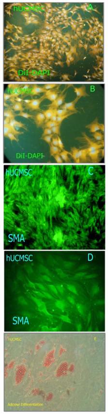
          Clinical studies of treating traumatic brain injury (TBI) with autologous adult stem cells led us to examine the impression of a combination therapy. This was performed by intravenous injection of human umbilical cord matrix stem cell (hUCMSC-Wharton ,s jelly stem cell) with bone marrow cell mobilized by granulocytecolony stimulating factor (G-CSF) in rats injured with cortical compact device.

          Materials and Methods

          Adult male Wistar rats (n= 50) were injured with controlled cortical impact device and divided into five groups. All injections were performed 1 day after injury into the tail veins of rats. Neurological functional evaluation of animals was performed before and after injury using modified neurological severity scores (mNSS). Animals were sacrificed 42 days after TBI and brain sections were stained by Brdu immunohistochemistry.

          Results

          Statistically significant improvement in functional outcome was observed in treatment groups when compared with control ( P< 0.01). mNSS showed no significant differences among the hUCMSC and G-CSF treated groups at any time point (end of trial). Rats with hUCMSC + G-CSF treatment had a significant improvement on mNSS at 5 and 6 week compared to other treatment group ( P< 0.01).

          Conclusion

          Histological analysis in G-CSF+ hUCMSC treated traumatic rats exhibited significant increase in numbers of Brdu immunoreactive cells in their traumatic core compared with other labeled group.

          Related collections

          Most cited references32

          • Record: found
          • Abstract: found
          • Article: not found

          Bone marrow as a potential source of hepatic oval cells.

          Bone marrow stem cells develop into hematopoietic and mesenchymal lineages but have not been known to participate in production of hepatocytes, biliary cells, or oval cells during liver regeneration. Cross-sex or cross-strain bone marrow and whole liver transplantation were used to trace the origin of the repopulating liver cells. Transplanted rats were treated with 2-acetylaminofluorene, to block hepatocyte proliferation, and then hepatic injury, to induce oval cell proliferation. Markers for Y chromosome, dipeptidyl peptidase IV enzyme, and L21-6 antigen were used to identify liver cells of bone marrow origin. From these cells, a proportion of the regenerated hepatic cells were shown to be donor-derived. Thus, a stem cell associated with the bone marrow has epithelial cell lineage capability.
            Bookmark
            • Record: found
            • Abstract: found
            • Article: not found

            A controlled cortical impact model of traumatic brain injury in the rat.

            Controlled cortical impact models produce brain injury by using a pneumatic impactor to impact exposed brain. This study systematically examined the effects of varying magnitudes of controlled cortical impact to the rat brain on neurological, cardiovascular, and histopathological variables. As the magnitude of injury increased, the duration of suppression of somatomotor reflexes and the duration of chronic vestibular motor deficits increased. The blood pressure response was observed to depend on injury levels; a moderate injury level produced a hypotensive response while a high injury level produced an immediate brief hypertensive response followed by hypotension. Low injury levels produced no significant macroscopic or microscopic change, but higher injury levels produced cortical contusion and intraparenchymal hemorrhage which, with increasing survival time, evolved into necrotic changes and cavitation underlying the injury site. Also with high levels of injury, axonal injury was found throughout the brain-stem with the greatest concentration of injured axons occurring in the cerebellar peduncles and pontomedullary junction. These data demonstrate that controlled cortical impact in the rat reproduces many of the features observed in other experimental animal models. This model allows independent control of many mechanical loading parameters associated with traumatic brain injury. The controlled cortical impact rat model should be an effective experimental tool to investigators of traumatic brain injury.
              Bookmark
              • Record: found
              • Abstract: found
              • Article: not found

              Intravenous administration of human bone marrow stromal cells induces angiogenesis in the ischemic boundary zone after stroke in rats.

              We tested the hypothesis that intravenous infusion of human bone marrow stromal cells (hMSCs) promotes vascular endothelial growth factor (VEGF) secretion, VEGF receptor 2 (VEGFR2) expression and angiogenesis in the ischemic boundary zone (IBZ) after stroke. hMSCs (1x10(6)) were intravenously injected into rats 24 hours after middle cerebral artery occlusion (MCAo). Laser scanning confocal microscopy (LSCM), immunohistochemistry and ELISA were performed to assay angiogenesis and levels of human and rat VEGF in the host brain, respectively. In addition, capillary-like tube formation was measured using mouse brain-derived endothelial cells (MBDECs). Morphological and three dimensional image analyses revealed significant (P<0.05) increases in numbers of enlarged and thin walled blood vessels and numbers of newly formed capillaries at the boundary of the ischemic lesion in rats (n=12) treated with hMSCs compared with numbers in rats (n=12) treated with PBS. ELISA measurements showed that treatment with hMSCs significantly (P<0.05) raised endogenous rat VEGF levels in the IBZ from 10.5+/-1.7 ng/mL in the control group to 17.5+/-1.6 ng/mL in the hMSC-treated group. In addition, treatment with hMSCs increased endogenous VEGFR2 immunoreactivity. In vitro, when MBDECs were incubated with the supernatant obtained from cultured hMSCs, capillary-like tube formation was significantly (P<0.01) induced. However, hMSC-induced capillary-like tube formation was significantly (P<0.01) inhibited when the endothelial cells were incubated with the supernatant from hMSCs in the presence of a neutralizing anti-VEGFR2. These data suggest that treatment of stroke with hMSCs enhances angiogenesis in the host brain and hMSC-enhanced angiogenesis is mediated by increases in levels of endogenous rat VEGF and VEGFR2.
                Bookmark

                Author and article information

                Journal
                Iran J Basic Med Sci
                Iran J Basic Med Sci
                IJBMS
                Iranian Journal of Basic Medical Sciences
                Mashhad University of Medical Sciences (Mashhad, Iran )
                2008-3866
                2008-3874
                Jul-Aug 2011
                : 14
                : 4
                : 327-339
                Affiliations
                [1 ] Cellular & Molecular Research Center, Tehran University of Medical Sciences, Tehran, Iran
                [2 ] Department of Anatomy, Tehran University of Medical Science, Tehran, Iran
                [3 ] Department of Biotechnology and Bioscience, Milano-Bicocca University, Milan, Italy
                [4 ] Department of Medical Physics, Tehran University of Medical Sciences, Tehran, Iran
                [5 ] Department of Immunology, Tehran University of Medical Sciences, Tehran, Iran
                [6 ] Department of physiology, Tehran University of Medical Sciences, Tehran, Iran
                Author notes
                [* ]Corresponding author: Tel/Fax: +98-21-88058689; email:mohsen136051@yahoo.com
                Article
                IJBMS-14-327
                3586827
                23492840
                4be80cf3-0a9c-425d-98fd-93813c0e4a1f
                © 2011: Iranian Journal of Basic Medical Sciences

                This is an Open Access article distributed under the terms of the Creative Commons Attribution License, ( http://creativecommons.org/licenses/by/3.0/) which permits unrestricted use, distribution, and reproduction in any medium, provided the original work is properly cited.

                History
                : 23 July 2010
                : 3 January 2011
                Categories
                original Article

                combine therapy,g-csf,stem cell,tbi,wharton,s jelly
                combine therapy, g-csf, stem cell, tbi, wharton, s jelly

                Comments

                Comment on this article