2
views
0
recommends
+1 Recommend
0 collections
    0
    shares
      • Record: found
      • Abstract: found
      • Article: found
      Is Open Access

      Emerging Roles of Sympathetic Nerves and Inflammation in Perivascular Adipose Tissue

      research-article

      Read this article at

      Bookmark
          There is no author summary for this article yet. Authors can add summaries to their articles on ScienceOpen to make them more accessible to a non-specialist audience.

          Abstract

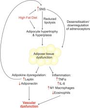
          Perivascular adipose tissue (PVAT) is no longer recognised as simply a structural support for the vasculature, and we now know that PVAT releases vasoactive factors which modulate vascular function. Since the discovery of this function in 1991, PVAT research is rapidly growing and the importance of PVAT function in disease is becoming increasingly clear. Obesity is associated with a plethora of vascular conditions; therefore, the study of adipocytes and their effects on the vasculature is vital. PVAT contains an adrenergic system including nerves, adrenoceptors and transporters. In obesity, the autonomic nervous system is dysfunctional; therefore, sympathetic innervation of PVAT may be the key mechanistic link between increased adiposity and vascular disease. In addition, not all obese people develop vascular disease, but a common feature amongst those that do appears to be the inflammatory cell population in PVAT. This review will discuss what is known about sympathetic innervation of PVAT, and the links between nerve activation and inflammation in obesity. In addition, we will examine the therapeutic potential of exercise in sympathetic stimulation of adipose tissue.

          Related collections

          Most cited references150

          • Record: found
          • Abstract: found
          • Article: not found

          Plasma adiponectin levels and risk of myocardial infarction in men.

          Adiponectin, a recently discovered adipocyte-derived peptide, is involved in the regulation of insulin sensitivity and lipid oxidation and, purportedly, in the development of atherosclerosis and coronary heart disease in humans. To assess prospectively whether plasma adiponectin concentrations are associated with risk of myocardial infarction (MI). Nested case-control study among 18 225 male participants of the Health Professionals Follow-up Study aged 40 to 75 years who were free of diagnosed cardiovascular disease at the time of blood draw (1993-1995). During 6 years of follow-up through January 31, 2000, 266 men subsequently developed nonfatal MI or fatal coronary heart disease. Using risk set sampling, controls were selected in a 2:1 ratio matched for age, date of blood draw, and smoking status (n = 532). Incidence of nonfatal MI and fatal coronary heart disease by adiponectin level. After adjustment for matched variables, participants in the highest compared with the lowest quintile of adiponectin levels had a significantly decreased risk of MI (relative risk [RR], 0.39; 95% confidence interval [CI], 0.23-0.64; P for trend <.001). Additional adjustment for family history of MI, body mass index, alcohol consumption, physical activity, and history of diabetes and hypertension did not substantively affect this relationship (RR, 0.41; 95% CI, 0.24-0.70; P for trend <.001). Further adjustment for hemoglobin A1c or C-reactive protein levels also had little impact, but additional adjustment for low- and high-density lipoprotein cholesterol levels modestly attenuated this association (RR, 0.56; 95% CI, 0.32-0.99; P for trend =.02). High plasma adiponectin concentrations are associated with lower risk of MI in men. This relationship can be only partly explained by differences in blood lipids and is independent of inflammation and glycemic status.
            Bookmark
            • Record: found
            • Abstract: found
            • Article: not found

            Physical activity and reduced risk of cardiovascular events: potential mediating mechanisms.

            Higher levels of physical activity are associated with fewer cardiovascular disease (CVD) events. Although the precise mechanisms underlying this inverse association are unclear, differences in several cardiovascular risk factors may mediate this effect. In a prospective study of 27,055 apparently healthy women, we measured baseline levels of hemoglobin A1c, traditional lipids (total, low-density lipoprotein, and high-density lipoprotein cholesterol), novel lipids [lipoprotein(a) and apolipoprotein A1 and B-100], creatinine, homocysteine, and inflammatory/hemostatic biomarkers (high-sensitivity C-reactive protein, fibrinogen, soluble intracellular adhesion molecule-1) and used women's self-reported physical activity, weight, height, hypertension, and diabetes. Mean follow-up was 10.9+/-1.6 years, and 979 incident CVD events occurred. The risk of CVD decreased linearly with higher levels of activity (P for linear trend or = 1500 kcal/wk of 27%, 32%, and 41%, respectively. Differences in known risk factors explained a large proportion (59.0%) of the observed inverse association. When sets of risk factors were examined, inflammatory/hemostatic biomarkers made the largest contribution to lower risk (32.6%), followed by blood pressure (27.1%). Novel lipids contributed less to CVD risk reduction compared with traditional lipids (15.5% and 19.1%, respectively). Smaller contributions were attributed to body mass index (10.1%) and hemoglobin A1c/diabetes (8.9%), whereas homocysteine and creatinine had negligible effects (< 1%). The inverse association between physical activity and CVD risk is mediated in substantial part by known risk factors, particularly inflammatory/hemostatic factors and blood pressure.
              Bookmark
              • Record: found
              • Abstract: found
              • Article: not found

              In vivo identification of bipotential adipocyte progenitors recruited by β3-adrenoceptor activation and high-fat feeding.

              Nutritional and pharmacological stimuli can dramatically alter the cellular phenotypes in white adipose tissue (WAT). Utilizing genetic lineage tracing techniques, we demonstrate that brown adipocytes (BA) that are induced by β3-adrenergic receptor activation in abdominal WAT arise from the proliferation and differentiation of cells expressing platelet-derived growth factor receptor alpha (PDGFRα), CD34, and Sca-1 (PDGFRα(+) cells). PDGFRα(+) cells have a unique morphology in which extended processes contact multiple cells in the tissue microenvironment. Surprisingly, these cells also give rise to white adipocytes (WA) that can comprise up to 25% of total fat cells in abdominal fat pads following 8 weeks of high-fat feeding. Isolated PDGFRα(+) cells differentiated into both BA and WA in vitro and generated WA after transplantation in vivo. The identification of PDGFRα(+) cells as bipotential adipocyte progenitors will enable further investigation of mechanisms that promote therapeutic cellular remodeling in adult WAT. Copyright © 2012 Elsevier Inc. All rights reserved.
                Bookmark

                Author and article information

                Contributors
                +44 161 275 1229 , sophie.saxton@manchester.ac.uk
                Journal
                Cardiovasc Drugs Ther
                Cardiovasc Drugs Ther
                Cardiovascular Drugs and Therapy
                Springer US (New York )
                0920-3206
                1573-7241
                12 February 2019
                12 February 2019
                2019
                : 33
                : 2
                : 245-259
                Affiliations
                [1 ]ISNI 0000000121662407, GRID grid.5379.8, Division of Cardiovascular Sciences, Manchester Academic Health Science Centre, , University of Manchester, ; Core Technology Facility (3rd floor), 46 Grafton Street, M13 9NT Manchester, UK
                [2 ]ISNI 0000 0004 0460 5971, GRID grid.8752.8, School of Environment and Life Sciences, , University of Salford, ; Manchester, UK
                Author information
                http://orcid.org/0000-0002-3067-1652
                Article
                6862
                10.1007/s10557-019-06862-4
                6509065
                30747398
                4bfa26ad-f0ef-459a-ab84-32893951ad1f
                © The Author(s) 2019

                OpenAccessThis article is distributed under the terms of the Creative Commons Attribution 4.0 International License (http://creativecommons.org/licenses/by/4.0/), which permits unrestricted use, distribution, and reproduction in any medium, provided you give appropriate credit to the original author(s) and the source, provide a link to the Creative Commons license, and indicate if changes were made.

                History
                Funding
                Funded by: FundRef http://dx.doi.org/10.13039/501100000274, British Heart Foundation;
                Award ID: PG/16/52/32229
                Award Recipient :
                Categories
                Original Article
                Custom metadata
                © Springer Science+Business Media, LLC, part of Springer Nature 2019

                Cardiovascular Medicine
                adipose tissue,inflammation,obesity,sympathetic nerves,exercise
                Cardiovascular Medicine
                adipose tissue, inflammation, obesity, sympathetic nerves, exercise

                Comments

                Comment on this article