1
views
0
recommends
+1 Recommend
0 collections
    0
    shares
      • Record: found
      • Abstract: found
      • Article: found
      Is Open Access

      Invasive Corridor of Clivus Extension in Pituitary Adenoma: Bony Anatomic Consideration, Surgical Outcome and Technical Nuances

      research-article

      Read this article at

      Bookmark
          There is no author summary for this article yet. Authors can add summaries to their articles on ScienceOpen to make them more accessible to a non-specialist audience.

          Abstract

          Background

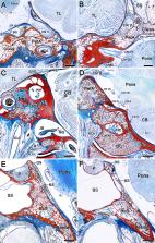
          It is well known that the clivus is composed of abundant cancellous bone and is often invaded by pituitary adenoma (PA), but the range of these cancellous bone corridors is unknown. In addition, we found that PA with clivus invasion is sometimes accompanied by petrous apex invasion, so we speculated that the petrous apex tumor originated from the clivus cancellous bone corridor. The aim of this study was to test this hypothesis by investigating the bony anatomy associated with PA with clival invasion and its clinical significance.

          Methods

          Twenty-two cadaveric heads were used in the anatomical study to research the bony architecture of the clivus and petrous apex, including six injected specimens for microsurgical dissection and sixteen cadavers for epoxy sheet plastination. The surgical videos and outcomes of PA with clival invasion in our single center were also retrospectively reviewed.

          Results

          The hypoglossal canal and internal acoustic meatus are composed of bone canals surrounded by cortical bone. The cancellous corridor within clivus starts from the sellar or sphenoid sinus floor and extends downward, bypassing the hypoglossal canal and finally reaching the occipital condyle and the medial edge of the jugular foramen. Interestingly, we found that the cancellous bone of the clivus was connected with that of the petrous apex through petroclival fissure extending to the medial margin of the internal acoustic meatus instead of a separating cortical bone between them as it should be. It is satisfactory that the anatomical outcomes of the cancellous corridor and the path of PA with clival invasion observed intraoperatively are completely consistent. In the retrospective cohort of 49 PA patients, the clival component was completely resected in 44 (89.8%), and only five (10.2%) patients in the early-stage had partial residual cases in the inferior clivus.

          Conclusion

          The petrous apex invasion of PA is caused by the tumor invading the clivus and crossing the petroclival fissure along the cancellous bone corridor. PA invade the clivus along the cancellous bone corridor and can also cross the hypoglossal canal to the occipital condyle. This clival invasion pattern presented here deepens our understanding of the invasive characteristics of PA.

          Related collections

          Most cited references31

          • Record: found
          • Abstract: found
          • Article: not found

          Invasion of the cavernous sinus space in pituitary adenomas: endoscopic verification and its correlation with an MRI-based classification.

          OBJECT An important prognostic factor for the surgical outcome and recurrence of a pituitary adenoma is its invasiveness into parasellar tissue, particularly into the space of the cavernous sinus (CS). The aims of this study were to reevaluate the existing parasellar classifications using an endoscopic technique and to evaluate the clinical and radiological outcomes associated with each grade. METHODS The authors investigated 137 pituitary macroadenomas classified radiologically at least on one side as Grade 1 or higher (parasellar extension) and correlated the surgical findings using an endoscopic technique, with special reference to the invasiveness of the tumor into the CS. In each case, postoperative MRI was performed to evaluate the gross-total resection (GTR) rate and the rate of endocrinological remission (ER) in functioning adenomas. RESULTS The authors found a 16% rate of CS invasion during surgery for these macroadenomas. Adenomas radiologically classified as Grade 1 were found to be invasive in 1.5%, and the GTR/ER rate was 83%/88%. For Grade 2 adenomas, the rate of invasion was 9.9%, and the GTR/ER rate was 71%/60%. For Grade 3 adenomas, the rate of invasion was 37.9%, and the GTR/ER rate was 75%/33%. When the superior compartment of the CS (Grade 3A) was involved, the authors found a rate of invasion that was lower (p < 0.001) than that when the inferior compartment was involved (Grade 3B). The rate of invasion in Grade 3A adenomas was 26.5% with a GTR/ER rate of 85%/67%, whereas for Grade 3B adenomas, the rate of surgically observed invasion was 70.6% with a GTR/ER rate of 64%/0%. All of the Grade 4 adenomas were invasive, and the GTR/ER rate was 0%. A comparison of microscopic and endoscopic techniques revealed no difference in adenomas with Grade 1 or 4 parasellar extension. In Grade 2 adenomas, however, the CS was found by the endoscopic technique to be invaded in 9.9% and by microscopic evaluation to be invaded in 88% (p < 0.001); in Grade 3 adenomas, the difference was 37.9% versus 86%, respectively (p = 0.002). Grade 4 adenomas had a statistically significant lower rate of GTR than those of all the other grades. In case of ER only, Grade 1 adenomas had a statistically significant higher rate of remission than did Grade 3B and Grade 4 adenomas. CONCLUSIONS The proposed classification proved that with increasing grades, the likelihood of surgically observed invasion rises and the chance of GTR and ER decreases. The direct endoscopic view confirmed the low rate of invasion of Grade 1 adenomas but showed significantly lower rates of invasion in Grade 2 and 3 adenomas than those previously found using the microscopic technique. In cases in which the intracavernous internal carotid artery was encased (Grade 4), all the adenomas were invasive and the GTR/ER rate was 0%/0%. The authors suggest the addition of Grades 3A and 3B to distinguish the strikingly different outcomes of adenomas invading the superior CS compartments and those invading the inferior CS compartments.
            Bookmark
            • Record: found
            • Abstract: found
            • Article: not found

            The long-term significance of microscopic dural invasion in 354 patients with pituitary adenomas treated with transsphenoidal surgery.

            Pituitary adenomas are considered benign tumors; however, they may infiltrate surrounding tissues including the dura mater. In this paper the authors analyze the clinical significance of microscopically confirmed dural invasion by comparing a range of variables (age and sex of patients, adenoma type, adenoma size on magnetic resonance [MR] images, remission, residual pituitary disease, recurrence, survival, and disease-free interval after surgery) between patients with noninvasive adenomas and those with invasive ones. Between 1992 and 1997 dural specimens were obtained in 354 patients with pituitary adenomas who underwent transsphenoidal surgery performed by the senior author (E.R.L.). Dural specimens were examined using routine histological methods and assessed for invasion by pituitary adenoma tissue. The dura was invaded by the pituitary adenoma in 161 patients (45.5%), and in 192 patients (54.5%) no evidence of dural invasion was found. Dural invasion was present significantly more frequently in the repeated surgery group (69%, 55 patients) than in the primary transsphenoidal surgery group (41%, 291 patients). The mean age of patients undergoing primary transsphenoidal surgery was significantly older in cases of invasive adenomas (50 years) compared with cases of noninvasive adenomas (43 years), and these age differences also correlated with adenoma size. Women tend to develop clinically evident, smaller adenomas at a younger age than men. Of the patients with pituitary adenomas that were 20 mm or smaller, 117 (76%) of 154 were women, whereas of the patients with adenomas that were larger than 20 mm, 74 (54%) of 137 were men. The frequency of dural invasion increased with increasing size of the pituitary adenoma as measured on MR images. In 291 patients who underwent primary pituitary surgery, the frequency of dural invasion according to adenoma size was 24% ( 10 to 20 to 40 mm). In patients who underwent primary transsphenoidal surgery, dural invasion was present in more than 50% of those with nonfunctioning adenomas and in 30 to 35% of patients with endocrinologically active adenomas. The mean diameter of the gonadotrophic adenomas and null-cell adenomas was significantly larger than that of each of the endocrinologically active adenomas. In 58 (20%) of 291 patients who underwent primary pituitary surgery there was residual pituitary disease postsurgery, and 20% of this subset of patients showed clinical improvement to such an extent that no further management was recommended. After pituitary surgery, residual tumor tissue was demonstrable significantly more frequently in patients with invasive adenomas than in those with noninvasive adenomas. Recurrences after initial remission (cure) of pituitary disease occurred in 18 (8.8%) of 205 patients between 2 and 79 months after primary pituitary surgery (median 25 months). The recurrence rate was not related to dural invasion in a consistent or significant fashion. Seven patients died between 14 and 79 months after pituitary surgery and all had invasive adenomas identified on gross observation at surgery and on microscopy. The survival rate was slightly but significantly decreased for patients with invasive adenomas (91%) compared with patients with noninvasive adenomas (100%) at 6 years postsurgery. The principal significance of dural invasion by pituitary adenoma is the persistence of tumor tissue after transsphenoidal surgery (incomplete adenomectomy; 20% in primary pituitary tumor resections). The increase in adenoma size with time and the concurrent development of dural invasion are the major factors that determine an incomplete adenomectomy. When the adenoma remains restricted to the sellar compartment or shows only moderate suprasellar extension, dural invasion may not yet have developed and conditions for complete selective adenomectomy are improved.
              Bookmark
              • Record: found
              • Abstract: found
              • Article: not found

              Expanded endonasal approach: fully endoscopic, completely transnasal approach to the middle third of the clivus, petrous bone, middle cranial fossa, and infratemporal fossa.

              The middle third of the clivus and the region around the petrous internal carotid artery (ICA) is a difficult area of the skull base in terms of access. This is a deep area rich with critical neurovascular structures, which is often host to typical skull base diseases. Expanded endoscopic endonasal approaches offer a potential option for accessing this difficult region. The objective of this paper was to establish the clinical feasibility of gaining access to the paraclival space in the region of the middle third of the clivus, to provide a practical modular and clinically applicable classification, and to describe the relevant critical surgical anatomy for each module. The anatomical organization of the region around the petrous ICA, cavernous sinus, and middle clivus is presented, with approaches divided into zones. In an accompanying paper in this issue by Cavallo, et al., the anatomy of the pterygopalatine fossa is presented; this was observed through cadaveric dissection for which an expanded endonasal approach was used. In the current paper the authors translate the aforementioned anatomical study to provide a clinically applicable categorization of the endonasal approach to the region around the petrous ICA. A series of zones inferior and superior to the petrous ICA are described, with an illustrative case presented for each region. The expanded endonasal approach is a feasible approach to the middle third of the clivus, petrous ICA, cavernous sinus, and medial infratemporal fossa in cases in which the lesion is located centrally, with neurovascular structures displaced laterally.
                Bookmark

                Author and article information

                Contributors
                Journal
                Front Oncol
                Front Oncol
                Front. Oncol.
                Frontiers in Oncology
                Frontiers Media S.A.
                2234-943X
                25 June 2021
                2021
                : 11
                : 689943
                Affiliations
                [1] 1 Department of Neurosurgery, The First Affiliated Hospital of Nanchang University , Nanchang, China
                [2] 2 Department of Anatomy, School of Basic Medical Sciences, Anhui Medical University , Hefei, China
                [3] 3 Department of Neurosurgery, Shanghai East Hospital, Tongji University School of Medicine , Shanghai, China
                Author notes

                Edited by: Xicai Sun, Fudan University, China

                Reviewed by: Ye Gu, Fudan University, China; Tomasz Dziedzic, Medical University of Warsaw, Poland; Tao Xie, Fudan University, China

                *Correspondence: Tao Hong, ht2000@ 123456vip.sina.com ; Liang Liang, liangliang@ 123456ahmu.edu.cn

                †These authors have contributed equally to this work

                This article was submitted to Surgical Oncology, a section of the journal Frontiers in Oncology

                Article
                10.3389/fonc.2021.689943
                8270656
                34249739
                502a0061-9dde-4521-974d-e8c5a6841cf8
                Copyright © 2021 Wu, Ding, Yang, Chu, Xie, Bao, Wu, Yang, Zhou, Li, Li, Tang, Xiao, Zhong, Liang and Hong

                This is an open-access article distributed under the terms of the Creative Commons Attribution License (CC BY). The use, distribution or reproduction in other forums is permitted, provided the original author(s) and the copyright owner(s) are credited and that the original publication in this journal is cited, in accordance with accepted academic practice. No use, distribution or reproduction is permitted which does not comply with these terms.

                History
                : 01 April 2021
                : 28 May 2021
                Page count
                Figures: 6, Tables: 1, Equations: 0, References: 31, Pages: 11, Words: 5037
                Categories
                Oncology
                Original Research

                Oncology & Radiotherapy
                pituitary adenoma,clival invasion,endonasal endoscopic approach,epoxy sheet plastination,corridor,cancellous bone,anatomy

                Comments

                Comment on this article