27
views
0
recommends
+1 Recommend
0 collections
    0
    shares
      • Record: found
      • Abstract: found
      • Article: found
      Is Open Access

      MicroRNA-133 overexpression promotes the therapeutic efficacy of mesenchymal stem cells on acute myocardial infarction

      research-article

      Read this article at

      Bookmark
          There is no author summary for this article yet. Authors can add summaries to their articles on ScienceOpen to make them more accessible to a non-specialist audience.

          Abstract

          Background

          Our study aim was to evaluate the therapeutic efficacy and mechanisms of miR-133-overexpressing mesenchymal stem cells (MSCs) on acute myocardial infarction.

          Methods

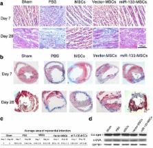
          Rat MSCs were isolated and purified by whole bone marrow adherent culturing. After transfection with the agomir or antagomir of miR-133, MSCs were collected for assay of cell vitality, apoptosis, and cell cycle progression. At the same time, exosomes were isolated from the supernatant to analyze the paracrine miR-133. For in-vivo studies, constitutive activation of miR-133 in MSCs was achieved by lentivirus-mediated miR-133 overexpression. A rat myocardial infarction model was created by ligating the left anterior descending coronary artery, while control MSCs (vector-MSCs) or miR-133-overexpressed MSCs (miR-133-MSCs) were injected into the zone around the myocardial infarction. Subsequently, myocardial function was evaluated by echocardiography on days 7 and 28 post infarction. Finally the infarcted hearts were collected on days 7 and 28 for myocardial infarct size measurement and detection of snail 1 expression.

          Results

          Hypoxia-induced apoptosis of MSCs obviously reduced, along with enhanced expression of total poly ADP-ribose polymerase protein, after miR-133 agomir transfection, while the apoptosis rate increased in MSCs transfected with miR-133 antagomir. However, no change in cell viability and cell-cycle distribution was observed in control, miR-133-overexpressed, and miR-133-interfered MSCs. Importantly, rats transplanted with miR-133-MSCs displayed more improved cardiac function after acute myocardial infarction, compared with those that received vector-MSC injection. Further studies indicated that cardiac expression of snail 1 was significantly repressed by adjacent miR-133-overexpressing MSCs, and both the inflammatory level and the infarct size decreased in miR-133-MSC-injected rat hearts.

          Conclusions

          miR-133-MSCs obviously improved cardiac function in a rat model of myocardial infarction. Transplantation of miR-133-overexpressing MSCs provides an effective strategy for cardiac repair and modulation of cardiac-related diseases.

          Electronic supplementary material

          The online version of this article (doi:10.1186/s13287-017-0722-z) contains supplementary material, which is available to authorized users.

          Related collections

          Most cited references27

          • Record: found
          • Abstract: not found
          • Article: not found

          Micromanagers of gene expression: the potentially widespread influence of metazoan microRNAs.

            Bookmark
            • Record: found
            • Abstract: found
            • Article: not found

            miR-133 and miR-30 regulate connective tissue growth factor: implications for a role of microRNAs in myocardial matrix remodeling.

            The myocardium of the failing heart undergoes a number of structural alterations, most notably hypertrophy of cardiac myocytes and an increase in extracellular matrix proteins, often seen as primary fibrosis. Connective tissue growth factor (CTGF) is a key molecule in the process of fibrosis and therefore seems an attractive therapeutic target. Regulation of CTGF expression at the promoter level has been studied extensively, but it is unknown how CTGF transcripts are regulated at the posttranscriptional level. Here we provide several lines of evidence to show that CTGF is importantly regulated by 2 major cardiac microRNAs (miRNAs), miR-133 and miR-30. First, the expression of both miRNAs was inversely related to the amount of CTGF in 2 rodent models of heart disease and in human pathological left ventricular hypertrophy. Second, in cultured cardiomyocytes and fibroblasts, knockdown of these miRNAs increased CTGF levels. Third, overexpression of miR-133 or miR-30c decreased CTGF levels, which was accompanied by decreased production of collagens. Fourth, we show that CTGF is a direct target of these miRNAs, because they directly interact with the 3' untranslated region of CTGF. Taken together, our results indicate that miR-133 and miR-30 importantly limit the production of CTGF. We also provide evidence that the decrease of these 2 miRNAs in pathological left ventricular hypertrophy allows CTGF levels to increase, which contributes to collagen synthesis. In conclusion, our results show that both miR-133 and miR-30 directly downregulate CTGF, a key profibrotic protein, and thereby establish an important role for these miRNAs in the control of structural changes in the extracellular matrix of the myocardium.
              Bookmark
              • Record: found
              • Abstract: found
              • Article: not found

              The muscle-specific microRNAs miR-1 and miR-133 produce opposing effects on apoptosis by targeting HSP60, HSP70 and caspase-9 in cardiomyocytes.

              The microRNAs miR-1 and miR-133 are preferentially expressed in cardiac and skeletal muscles and have been shown to regulate differentiation and proliferation of these cells. We report here a novel aspect of cellular function of miR-1 and miR-133 regulation of cardiomyocyte apoptosis. miR-1 and miR-133 produced opposing effects on apoptosis, induced by oxidative stress in H9c2 rat ventricular cells, with miR-1 being pro-apoptotic and miR-133 being anti-apoptotic. miR-1 level was significantly increased in response to oxidative stress. We identified single target sites for miR-1 only, in the 3'-untranslated regions of the HSP60 and HSP70 genes, and multiple putative target sites for miR-133 throughout the sequence of the caspase-9 gene. miR-1 reduced the levels of HSP60 and HSP70 proteins without changing their transcript levels, whereas miR-133 did not affect HSP60 and HSP70 expression at all. By contrast, miR-133 repressed caspase-9 expression at both the protein and mRNA levels. The post-transcriptional repression of HSP60 and HSP70 and caspase-9 was further confirmed by luciferase reporter experiments. Our results indicate that miR-1 and miR-133 are involved in regulating cell fate with increased miR-1 and/or decreased miR-133 levels favoring apoptosis and decreased miR-1 and/or miR-133 levels favoring survival. Post-transcriptional repression of HSP60 and HSP70 by miR-1 and of caspase-9 by miR-133 contributes significantly to their opposing actions.
                Bookmark

                Author and article information

                Contributors
                chenyueqiu-216@163.com
                906528529@qq.com
                chenweiqian0211@gmail.com
                1085815297@qq.com
                zhaozhenao@suda.edu.cn
                jjyang@suda.edu.cn
                18605122344@126.com
                leiwei@suda.edu.cn
                uuzyshen@aliyun.com
                Journal
                Stem Cell Res Ther
                Stem Cell Res Ther
                Stem Cell Research & Therapy
                BioMed Central (London )
                1757-6512
                25 November 2017
                25 November 2017
                2017
                : 8
                : 268
                Affiliations
                [1 ]ISNI 0000 0001 0198 0694, GRID grid.263761.7, Institute for Cardiovascular Science & Department of Cardiovascular Surgery of The First Affiliated Hospital, , Soochow University, ; 215007, 708 Renmin Rd, Bldg 1, Suzhou, China
                [2 ]Nantong First People’s Hospital, 226001, North Rd, Haier alley, Nantong, China
                Article
                722
                10.1186/s13287-017-0722-z
                5702098
                29178928
                51249255-dafd-4979-a9f3-7b77fb1226cd
                © The Author(s). 2017

                Open AccessThis article is distributed under the terms of the Creative Commons Attribution 4.0 International License ( http://creativecommons.org/licenses/by/4.0/), which permits unrestricted use, distribution, and reproduction in any medium, provided you give appropriate credit to the original author(s) and the source, provide a link to the Creative Commons license, and indicate if changes were made. The Creative Commons Public Domain Dedication waiver ( http://creativecommons.org/publicdomain/zero/1.0/) applies to the data made available in this article, unless otherwise stated.

                History
                : 31 July 2017
                : 5 November 2017
                : 7 November 2017
                Funding
                Funded by: National Natural Science Foundation (NSFC) of China
                Award ID: 81600218
                Award Recipient :
                Funded by: National Natural Science Foundation (NSFC) of China
                Award ID: 31401239
                Award Recipient :
                Funded by: Jiangsu clinical research center for cardiovascular surgery
                Funded by: National Clinical Key Specialty of cardiovascular surgery
                Funded by: FundRef http://dx.doi.org/10.13039/501100004608, Natural Science Foundation of Jiangsu Province;
                Award ID: BK20150320
                Award ID: BK20150321
                Award Recipient :
                Funded by: Suzhou Municipal Science and Technology Foundation
                Award ID: SYS201540
                Award ID: SYS201675
                Award Recipient :
                Funded by: Natural Science Fund for Colleges and Universities in Jiangsu Province
                Award ID: 14KJB180021
                Award Recipient :
                Funded by: Natural Science Fund for Colleges and Universities in Jiangsu Province
                Award ID: 15KJB180017
                Award Recipient :
                Categories
                Research
                Custom metadata
                © The Author(s) 2017

                Molecular medicine
                mir-133,mesenchymal stem cells,myocardial infarction
                Molecular medicine
                mir-133, mesenchymal stem cells, myocardial infarction

                Comments

                Comment on this article