0
views
0
recommends
+1 Recommend
0 collections
    0
    shares
      • Record: found
      • Abstract: found
      • Article: found
      Is Open Access

      Multispectral imaging of nailfold capillaries using light-emitting diode illumination

      research-article

      Read this article at

      Bookmark
          There is no author summary for this article yet. Authors can add summaries to their articles on ScienceOpen to make them more accessible to a non-specialist audience.

          Abstract.

          Significance

          The capillaries are the smallest blood vessels in the body, typically imaged using video capillaroscopy to aid diagnosis of connective tissue diseases, such as systemic sclerosis. Video capillaroscopy allows visualization of morphological changes in the nailfold capillaries but does not provide any physiological information about the blood contained within the capillary network. Extracting parameters such as hemoglobin oxygenation could increase sensitivity for diagnosis and measurement of microvascular disease progression.

          Aim

          To design, construct, and test a low-cost multispectral imaging (MSI) system using light-emitting diode (LED) illumination to assess relative hemoglobin oxygenation in the nailfold capillaries.

          Approach

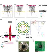
          An LED ring light was first designed and modeled. The ring light was fabricated using four commercially available LED colors and a custom-designed printed circuit board. The experimental system was characterized and results compared with the illumination model. A blood phantom with variable oxygenation was used to determine the feasibility of using the illumination-based MSI system for oximetry. Nailfold capillaries were then imaged in a healthy subject.

          Results

          The illumination modeling results were in close agreement with the constructed system. Imaging of the blood phantom demonstrated sensitivity to changing hemoglobin oxygenation, which was in line with the spectral modeling of reflection. The morphological properties of the volunteer capillaries were comparable to those measured in current gold standard systems.

          Conclusions

          LED-based illumination could be used as a low-cost approach to enable MSI of the nailfold capillaries to provide insight into the oxygenation of the blood contained within the capillary network.

          Related collections

          Most cited references46

          • Record: found
          • Abstract: found
          • Article: not found

          2013 classification criteria for systemic sclerosis: an American College of Rheumatology/European League against Rheumatism collaborative initiative.

          The 1980 American College of Rheumatology (ACR) classification criteria for systemic sclerosis (SSc) lack sensitivity for early SSc and limited cutaneous SSc. The present work, by a joint committee of the ACR and the European League Against Rheumatism (EULAR), was undertaken for the purpose of developing new classification criteria for SSc. Using consensus methods, 23 candidate items were arranged in a multicriteria additive point system with a threshold to classify cases as SSc. The classification system was reduced by clustering items and simplifying weights. The system was tested by 1) determining specificity and sensitivity in SSc cases and controls with scleroderma-like disorders, and 2) validating against the combined view of a group of experts on a set of cases with or without SSc. It was determined that skin thickening of the fingers extending proximal to the metacarpophalangeal joints is sufficient for the patient to be classified as having SSc; if that is not present, 7 additive items apply, with varying weights for each: skin thickening of the fingers, fingertip lesions, telangiectasia, abnormal nailfold capillaries, interstitial lung disease or pulmonary arterial hypertension, Raynaud's phenomenon, and SSc-related autoantibodies. Sensitivity and specificity in the validation sample were, respectively, 0.91 and 0.92 for the new classification criteria and 0.75 and 0.72 for the 1980 ACR classification criteria. All selected cases were classified in accordance with consensus-based expert opinion. All cases classified as SSc according to the 1980 ACR criteria were classified as SSc with the new criteria, and several additional cases were now considered to be SSc. The ACR/EULAR classification criteria for SSc performed better than the 1980 ACR criteria for SSc and should allow for more patients to be classified correctly as having the disease. Copyright © 2013 by the American College of Rheumatology.
            Bookmark
            • Record: found
            • Abstract: found
            • Article: found
            Is Open Access

            Globally optimal stitching of tiled 3D microscopic image acquisitions

            Motivation: Modern anatomical and developmental studies often require high-resolution imaging of large specimens in three dimensions (3D). Confocal microscopy produces high-resolution 3D images, but is limited by a relatively small field of view compared with the size of large biological specimens. Therefore, motorized stages that move the sample are used to create a tiled scan of the whole specimen. The physical coordinates provided by the microscope stage are not precise enough to allow direct reconstruction (Stitching) of the whole image from individual image stacks. Results: To optimally stitch a large collection of 3D confocal images, we developed a method that, based on the Fourier Shift Theorem, computes all possible translations between pairs of 3D images, yielding the best overlap in terms of the cross-correlation measure and subsequently finds the globally optimal configuration of the whole group of 3D images. This method avoids the propagation of errors by consecutive registration steps. Additionally, to compensate the brightness differences between tiles, we apply a smooth, non-linear intensity transition between the overlapping images. Our stitching approach is fast, works on 2D and 3D images, and for small image sets does not require prior knowledge about the tile configuration. Availability: The implementation of this method is available as an ImageJ plugin distributed as a part of the Fiji project ( F iji i s j ust I mageJ: http://pacific.mpi-cbg.de/). Contact: tomancak@mpi-cbg.de
              Bookmark
              • Record: found
              • Abstract: found
              • Article: found
              Is Open Access

              Photoacoustic tomography of blood oxygenation: A mini review

              Photoacoustic tomography (PAT) is a hybrid imaging modality that combines rich contrast of optical excitation and deep penetration of ultrasound detection. With its unique optical absorption contrast mechanism, PAT is inherently sensitive to the functional and molecular information of biological tissues, and thus has been widely used in preclinical and clinical studies. Among many functional capabilities of PAT, measuring blood oxygenation is arguably one of the most important applications, and has been widely performed in photoacoustic studies of brain functions, tumor hypoxia, wound healing, and cancer therapy. Yet, the complex optical conditions of biological tissues, especially the strong wavelength-dependent optical attenuation, have long hurdled the PAT measurement of blood oxygenation at depths beyond a few millimeters. A variety of PAT methods have been developed to improve the accuracy of blood oxygenation measurement, using novel laser illumination schemes, oxygen-sensitive fluorescent dyes, comprehensive mathematic models, or prior information provided by complementary imaging modalities. These novel methods have made exciting progress, while several challenges remain. This concise review aims to introduce the recent developments in photoacoustic blood oxygenation measurement, compare each method’s advantages and limitations, highlight their representative applications, and discuss the remaining challenges for future advances.
                Bookmark

                Author and article information

                Contributors
                Journal
                J Biomed Opt
                J Biomed Opt
                JBOPFO
                JBO
                Journal of Biomedical Optics
                Society of Photo-Optical Instrumentation Engineers
                1083-3668
                1560-2281
                12 December 2022
                December 2022
                12 December 2022
                : 27
                : 12
                : 126002
                Affiliations
                [a ]University of Cambridge , Department of Physics, Cavendish Laboratory, Cambridge, United Kingdom
                [b ]University of Cambridge , Cancer Research UK Cambridge Institute, Cambridge, United Kingdom
                [c ]University of Arizona , Wyant College of Optical Sciences, Tucson, Arizona, United States
                [d ]University of Manchester , Division of Cancer Sciences, Quantitative Biomedical Imaging Laboratory, Manchester, United Kingdom
                [e ]University of Manchester , NIHR Manchester Biomedical Research Centre, Manchester, United Kingdom
                Author notes
                [* ]Address all correspondence to Sarah E. Bohndiek, seb53@ 123456cam.ac.uk
                Author information
                https://orcid.org/0000-0003-3279-2674
                https://orcid.org/0000-0002-6911-0289
                https://orcid.org/0000-0002-6432-6515
                https://orcid.org/0000-0003-4727-2006
                https://orcid.org/0000-0001-9244-2882
                https://orcid.org/0000-0003-0371-8635
                Article
                JBO-220109GRR 220109GRR
                10.1117/1.JBO.27.12.126002
                9743620
                36519074
                5339eacc-6bfe-4179-be01-a67747ea67f9
                © 2022 The Authors

                Published by SPIE under a Creative Commons Attribution 4.0 International License. Distribution or reproduction of this work in whole or in part requires full attribution of the original publication, including its DOI.

                History
                : 22 May 2022
                : 9 November 2022
                Page count
                Figures: 6, Tables: 4, References: 46, Pages: 15
                Funding
                Funded by: General Sir John Monash Foundation
                Funded by: Cambridge Trust
                Funded by: EPSRC
                Award ID: EP/S022139/1
                Funded by: EPSRC
                Award ID: EP/R003599/1
                Funded by: Wellcome Trust
                Funded by: Cancer Research UK
                Award ID: C9545/A29580
                Categories
                Imaging
                Paper
                Custom metadata
                Taylor-Williams et al.: Multispectral imaging of nailfold capillaries using light-emitting diode illumination

                Biomedical engineering
                multispectral imaging,nailfold capillaroscopy,light-emitting diode (led),oximetry,hemoglobin,phantom,illumination modeling,spectral modeling

                Comments

                Comment on this article