- Record: found
- Abstract: found
- Article: found
Diagnosis and Management of Cardiovascular Disease in Advanced and End‐Stage Renal Disease
review-article
Navdeep K. Bhatti , MD
1 ,
Keyvan Karimi Galougahi , MD, PhD
1 ,
Yehuda Paz , MD
1 ,
Tamim Nazif , MD
1
,
4 ,
Jeffrey W. Moses , MD
1
,
4 ,
Martin B. Leon , MD
1
,
4 ,
Gregg W. Stone , MD
1
,
4 ,
Ajay J. Kirtane , MD
1
,
4 ,
Dimitri Karmpaliotis , MD
1
,
4 ,
Sabahat Bokhari , MD
1 ,
Mark A. Hardy , MD
3 ,
Geoffrey Dube , MD
2 ,
Sumit Mohan , MD
2 ,
Lloyd E. Ratner , MD, MPH
3 ,
David J. Cohen , MD
2 ,
Ziad A. Ali , MD, DPhil
1
,
4
,
04 August 2016
Read this article at
There is no author summary for this article yet. Authors can add summaries to their articles on ScienceOpen to make them more accessible to a non-specialist audience.
Abstract
Introduction
Chronic kidney disease (CKD) affects 13% of the US population.1 Although a significant
proportion of these patients progress to end‐stage renal disease (ESRD) requiring
renal replacement therapy (RRT)2 or renal transplantation, cardiovascular disease
remains the most common cause of mortality and accounts for 53% of all deaths with
a known cause in patients on dialysis.3 Critically, cardiovascular disease also remains
the leading cause of death after renal transplantation. Appropriate management of
cardiovascular disease in this very high‐risk population is of paramount importance.
Pathobiological processes that underpin the progression and severity of cardiovascular
disease in CKD include accelerated atherosclerosis and continuous reduction in left
ventricular (LV) function as renal function declines.1 While on hemodialysis, these
processes accelerate. Importantly, the risk of developing pulmonary hypertension (PH)
also rises proportionately to the duration of hemodialysis.4 In contrast to dialysis,
renal transplantation can help prevent the progression of pathological cardiovascular
processes. Renal transplantation can potentially reverse myocardial damage that is
thought to result from prolonged exposure to uremic toxins and improve LV systolic
function.5, 6, 7
In this review, we provide a contemporary overview of the pre‐ and perioperative cardiovascular
evaluation of patients with ESRD who are considered suitable candidates for renal
transplantation. In addition, we review the evidence‐based guidelines on optimal management
of cardiovascular disease in patients with advanced CKD with particular focus on coronary
artery disease (CAD), congestive heart failure (CHF), valvular disease, and PH. The
overall aim is to identify the subset of patients who may maximally benefit from renal
transplantation. Finally, we provide evidence‐based recommendations for diagnosis,
management, and application in clinical practice.
CAD in Patients With ESRD
CAD is highly prevalent in patients with ESRD largely because of the presence of comorbidities
such as hypertension, diabetes mellitus, dyslipidemia, obesity, and tobacco use.8
The incidence of CAD in patients initiating dialysis is up to 38%, with a relative
risk of 5‐ to 20‐fold that of the general population.9 The uremic environment may
also contribute to the higher prevalence and accelerated progression of CAD.1, 10
Moreover, atherosclerosis is an inflammatory process.11, 12 Patients with ESRD have
high levels of C‐reactive protein and proinflammatory cytokines,1, 10, 13, 14 which
predisposes them to plaque formation. Endothelial dysfunction and high oxidative stress
further drive atherosclerosis and are exacerbated in the setting of the activated
renin–angiotensin–aldosterone system in CKD and ESRD.1, 10, 13, 14 Moreover, therapies
for secondary prevention of CAD such as statins and angiotensin‐converting enzyme
(ACE) inhibitors may have diminished clinical benefit in ESRD.12, 15
Coronary plaques in patients with ESRD exhibit extensive heterotopic calcification.16
On computed tomography coronary angiography in young patients with ESRD, a disproportionate
incidence of high calcium scores is detected with the probability of coronary artery
calcification increasing with longer durations of dialysis.16 Calcification occurs
in smooth muscle cells in the media or in the neointima of atherosclerotic plaques,
contributing to vascular stiffness and death from CAD.17 In addition to increased
plaque complexity, the clinical presentation of CAD is also different. Patients with
advanced CKD are more likely to present with acute coronary syndrome as the first
manifestation of CAD, as opposed to angina in patients without renal disease.18
Noninvasive Imaging to Assess CAD
Many sets of guidelines aim to guide cardiovascular evaluation in renal transplantation
candidates, but there is no universal consensus on an optimal approach. The 2014 American
College of Cardiology (ACC) and American Heart Association (AHA) guidelines on perioperative
cardiovascular evaluation in the general population undergoing noncardiac surgery
do not recommend testing for asymptomatic patients with a functional capacity considered
to be moderate (defined as ≥4 metabolic equivalents).19 Testing in patients with poor
functional capacity (<4 metabolic equivalents) or unknown functional status is recommended
to be based on combined clinical and surgical risk factors, with noninvasive tests
performed for patients at elevated risk19. However, it is unclear whether these recommendations
should be applied to potential candidates for transplantation.20 A study of 204 candidates
for renal transplantation reported that 80% of patients with no active cardiac conditions
had a functional status of ≥4 metabolic equivalents,20, 21 which in part reflects
the relatively younger age of transplant candidates. Consequently, a functional status
of ≥4 metabolic equivalents is not a reliable predictor of CAD in this population.20,
21 Instead, the 2012 AHA/ACC scientific statement regarding cardiac evaluation in
renal transplantation candidates recommends that the decision to proceed with noninvasive
stress testing in patients with no active cardiac conditions should be based on the
presence of multiple risk factors for CAD most relevant to the transplantation population,
regardless of functional status. These risk factors include diabetes mellitus, prior
cardiovascular disease, >1 year on dialysis, left ventricular hypertrophy (LVH), age
>60 years, smoking, hypertension, and dyslipidemia.20, 22 Although the specific number
of risk factors to proceed with stress testing remains to be determined, the AHA/ACC
guidelines suggest the presence of ≥3 risk factors as a reasonable threshold for noninvasive
testing.20 Guidelines from the Kidney Disease Outcomes Quality Initiative (KDOQI)
recommend annual evaluation for CAD in diabetic patients on the waiting list for transplantation
if the initial evaluation for CAD at the start of dialysis is negative. In high‐risk
patients without diabetes mellitus on the transplantation waitlist (≥2 traditional
risk factors, known history of CAD, peripheral vascular disease, and LV ejection fraction
[LVEF] ≤40%), evaluation for CAD every 24 months is recommended. Patients on hemodialysis
with an LVEF ≤40% or those with new symptoms of concern regarding ischemic heart disease
are recommended to be evaluated continuously for CAD.23, 24
Noninvasive testing with electrocardiogram (ECG), transthoracic echocardiogram, pharmacological
stress echocardiography, and nuclear imaging (with single photon emission computed
tomography [SPECT] or cardiac positron emission tomography [PET]) are suggested as
the first steps in investigating for presence of CAD. Patients should have baseline
ECGs to evaluate for Q waves, ST‐T changes, T wave inversions, and left bundle branch
block, which previously have been shown to be predictive of CAD.25 Exercise ECG is
not recommended, given abnormal baseline ECGs and overall poor exercise tolerance
in this patient population. A baseline transthoracic echocardiogram performed at dry
weight is also important because it can help identify impaired LVEF and wall motion
abnormalities, which may be signs of prognostically significant CAD.26 A normal cardiac
stress test has a high negative predictive value for cardiovascular events27, 28 in
the perioperative and follow‐up periods, as shown in a study of renal transplant candidates
undergoing preoperative SPECT.29 A hybrid SPECT/computed tomography scan assesses
for ischemia and coronary artery calcification, which is highly prevalent in patients
with ESRD.30 Coronary artery calcium score, however, does not independently provide
significant incremental prognostic value in predicting mortality or nonfatal myocardial
infarction in ESRD.30 These findings may be explained by the differences in distribution
of calcium within the coronary artery in ESRD, as shown by intravascular imaging.31
Patients with ESRD have a higher prevalence of intimal calcium without greater lipid
arc or thin‐cap fibroatheroma, which are markers of vulnerable plaque.31
Presence of inducible ischemia on dobutamine stress echocardiogram (DSE) has been
shown to be predictive of future cardiac events and all‐cause mortality.27, 32 Although
the accuracy of dobutamine stress echocardiogram and SPECT in detecting obstructive
CAD (≥70% stenosis) in renal transplantation candidates was not statistically different
in a meta‐analysis,33 the presence of concentric and eccentric LVH, common in ESRD,
may affect the accuracy of dobutamine stress echocardiogram.34 PET imaging assesses
not only myocardial blood flow but also coronary flow reserve, which can provide additional
insights into early stages of atherosclerosis and microvascular dysfunction.35, 36
Recently, coronary flow reserve assessed by cardiac PET has been shown to provide
incremental risk stratification for cardiovascular and all‐cause mortality in patients
on dialysis, even in the absence of overt cardiovascular disease.36 For the highest
risk patients, PET may be advantageous because it has superior sensitivity for detecting
CAD.35, 36 In addition, PET exposes patients to far less radiation than SPECT, an
important consideration given the potential need for repeated stress testing during
the recipient waiting period. Overall, it is important to consider both the local
availability of these tests and the expertise in interpreting them when deciding which
test is best suited to evaluate for ischemia in this group of patients.
Coronary Angiography and Revascularization
Although coronary angiography is usually reserved for patients with evidence of ischemia
on noninvasive imaging to determine their need for preoperative revascularization,
it is also reasonable to consider coronary angiography in renal transplantation candidates
at high risk of CAD despite normal stress tests. It is important to identify prognostically
important CAD that may require revascularization prior to transplantation.37 Evidence
of atherosclerotic vascular disease involving other vascular beds, particularly peripheral
arterial disease, may help identify patients with advanced coronary atherosclerosis.38,
39, 40, 41 Since peripheral arterial disease is highly associated with CAD, it may
be reasonable to pursue left heart catheterization in patients with peripheral arterial
disease despite negative stress tests. Similarly, patients with cardiac autonomic
dysfunction, autonomic neuropathy, and retinopathy are at increased risk of CAD.42,
43, 44 The Detection of Ischemia in Asymptomatic Diabetics (DIAD) study found that
cardiac autonomic dysfunction was a major predictor of inducible ischemia.42 Furthermore,
some diabetic patients with retinopathy have been found to have reduced coronary flow
reserve and cardiovascular disease.43, 44 Taken together, renal transplant candidates
with diabetes mellitus as the primary etiology for CKD who have normal stress tests
may represent a particularly high‐risk group that should be considered for coronary
angiography, given their high pretest probability for CAD, as myocardial perfusion
imaging has a high false‐negative rate in this population.37
Revascularization With Coronary Artery Bypass Grafting Versus Percutaneous Coronary
Intervention
Observational studies in patients with ESRD who undergo revascularization with percutaneous
coronary intervention (PCI) or coronary artery bypass grafting (CABG) surgery have
shown similar long‐term outcomes.45, 46, 47, 48, 49 A recent retrospective analysis
of >13 000 patients with CKD treated with CABG or PCI revealed that in the first 3 months
after surgery, patients who underwent CABG had a higher risk of progression to ESRD
and a higher mortality rate compared with those who underwent PCI.50 This study used
more contemporary interventional approaches such as drug‐eluting stents (DESs) as
opposed to older generation bare metal stents (BMSs) which helped improve postprocedural
cardiovascular outcomes.50 After the first 6 months, however, CABG portended improved
survival. An observational study evaluating >21 000 patients with CKD and multivessel
CAD undergoing PCI or CABG revealed improved 5‐year survival rates in patients who
received CABG51; however, these results do not apply to patients with single‐ or double‐vessel
CAD. It is important to note that this study did not take into account LV systolic
dysfunction, which places patients at a higher risk of sustaining a cardiac event
in the perioperative and postoperative periods. It is possible that patients who underwent
PCI as opposed to CABG for multivessel disease had a high operative risk that precluded
surgical intervention. The KDOQI guidelines recommend CABG for significant left main
or 3‐vessel CAD.24
Although randomized controlled trials to determine the overall mortality benefit of
revascularization with PCI in patients with ESRD are lacking, nonrandomized data have
suggested that PCI can reduce mortality and lead to greater cardiac event–free survival
after transplantation.52 The ongoing ISCHEMIA‐CKD study (ClinicalTrials.gov identifier
NCT01985360) will provide critical data for evidence‐based management of CAD in patients
with CKD. A retrospective analysis of 1460 renal transplant candidates revealed that
patients who underwent preoperative coronary revascularization with PCI had significantly
improved 5‐year survival after renal transplantation compared with patients who were
medically managed.52 Despite similar indications for revascularization in patients
with ESRD, this high‐risk group of patients was significantly less likely to be revascularized
compared with patients with normal renal function,20, 53 in particular those patients
with CKD not yet requiring RRT, as contrast‐induced nephropathy can precipitate the
need for dialysis. To preserve renal function, our group has recently reported the
safety and feasibility of cardiac revascularization with PCI guided by intravascular
imaging and coronary physiology without utilizing radiocontrast in patients with advanced
CKD (stages 4–5).54 This strategy may be adapted in centers with expertise in intravascular
imaging and physiology and may lead to increased provision of PCI while protecting
against contrast‐induced nephropathy and need for RRT. Unfortunately, even in the
setting of acute myocardial infarction, patients with CKD are less likely to be revascularized
than patients with normal renal function,55 despite similar health‐related quality
of life compared with patients without CKD in the post–myocardial infarction period.
In addition, patients with CKD were less likely to be prescribed guideline‐recommended
therapy including aspirin, statins, and ACE inhibitors at discharge,55 which may contribute
to higher cardiovascular morbidity and mortality. Medical treatment aimed at improving
health status after acute myocardial infarction should focus on all patients and be
based on current guidelines, regardless of CKD status. The decision to pursue revascularization
(CABG or PCI) or to treat medically should be made after a multidisciplinary discussion
among interventional cardiologists, nephrologists, and cardiothoracic surgeons and
should be individualized to each patient, given the current lack of evidence to guide
therapy.56
DES Versus BMS
The choice of the types of stents used for PCI in patients with CKD and ESRD presents
a clinical dilemma. Compared with patients with normal renal function, restenosis
rates in patients with CKD and ESRD are significantly higher.51, 53, 57, 58, 59 Several
studies have shown that the implantation of a DES is associated with lower rates of
target vessel revascularization compared with patients who receive a BMS. Nevertheless,
the risk of restenosis with a DES in patients with ESRD compared with patients with
normal renal function is still higher.51, 53, 57, 58, 59 A randomized multicenter
study evaluating the efficacy of everolimus‐eluting stents versus BMSs of identical
size and implanted in the same patient showed a reduction in ischemia‐driven target
vessel revascularization in patients with CKD who received DESs.60 Nevertheless, it
is important to note that a BMS may be preferred in patients in whom renal transplantation
is planned within 6 to 12 months, such as those planned to receive living donor transplants,
to limit the duration of dual antiplatelet therapy (DAPT). More novel stents that
are polymer‐ and carrier‐free but are drug‐coated have been shown to be superior to
BMSs with respect to requiring target vessel revascularization on only 1 month of
DAPT.61 This type of stent confers the benefit of a DES with an improved safety profile
over a BMS, with the additional advantage of a short course of DAPT, which may be
ideal for patients with ESRD.61 Aggressive medical therapy is also an option, given
the high restenosis rates in patients with CKD and ESRD.62, 63, 64 These risks must
be weighed against the observed improved survival rate after PCI versus medical therapy
alone in patients with ESRD.65
Duration of DAPT
The 2014 ACC/AHA guidelines recommend that in patients undergoing urgent noncardiac
surgery performed <4 to 6 months after BMS or DES implantation, DAPT should be continued
unless the relative risk of bleeding outweighs the benefit of prevention of stent
thrombosis.19 Moreover, DAPT should be continued for at least 6 months in patients
with DESs if the risk of surgical delay is greater than the risk of DES thrombosis.
Importantly, the ACC/AHA guidelines also recommend that perioperative management of
DAPT should be discussed by a multidisciplinary team including the operating surgeon,
cardiologist, anesthesiologist, and patient to weigh the risks of bleeding and stent
thrombosis in an individualized fashion. Important factors to consider in perioperative
DAPT management are the type, number, and size of stents versus the risk of delaying
renal transplantation in favor of prolonging DAPT to prevent stent thrombosis. A newer
generation of DESs with enhanced biocompatibility and reduced thrombogenicity may
require only 1 to 6 months of DAPT depending on the type of stent, but more evidence
is needed.61, 66, 67 These considerations highlight the fact that DAPT should be tailored
to the individual patient. For example, patients requiring multivessel PCI with the
use of a DES for lesions involving coronary ostia, long lesions, bifurcation lesions,
chronic total occlusions, or for cases in which the minimal stent area achieved is
small, may benefit from a prolonged course of DAPT. In contrast, patients with high
bleeding risk or single‐vessel disease who undergo PCI with a DES for simpler coronary
lesions (AHA type A/B1) with a large minimal stent area achieved by imaging guidance
may require shorter duration of DAPT.68, 69 Moreover, it is important to note that
patients with CKD have less platelet inhibition by clopidogrel.70 Prasugrel and ticagrelor
have not been well studied in CKD, and ticagrelor has a relative contraindication
in patients with CKD. Data from a few recent clinical series show similar outcomes
of renal transplantation performed on DAPT soon after DES implantation. Patients on
DAPT or aspirin alone compared with patients not receiving antiplatelet therapy did
not have a statistically significant increase in bleeding risk, requirement for blood
transfusion, or reoperation.71, 72 These studies should be interpreted with caution
because the long‐term effects of increased periprocedural bleeding and the need for
transfusion on graft survival are lacking.
Effects of Renal Transplantation on Incidence of Acute Coronary Syndrome
Renal transplantation decreases the incidence of acute coronary syndrome compared
with maintenance dialysis.73, 74, 75 Renal transplantation is also independently associated
with a lower risk of acute coronary syndrome in patients with ESRD secondary to diabetes
mellitus compared with patients maintained on hemodialysis.74 Furthermore, transplantation
is associated with a 17% lower adjusted risk of acute coronary syndrome compared with
patients remaining on the waiting list for transplantation regardless of the etiology
of ESRD.74 In the posttransplantation period, the risk of ischemic heart disease persists,
albeit attenuated, compared with maintenance dialysis; acute graft failure, LVH, and
traditional cardiovascular risk factors are the major predictors of ischemic events.23
Furthermore, immunosuppressive therapy with calcineurin inhibitors and steroids required
in transplanted patients can induce diabetes mellitus and worsen glycemic control,
hypertension, and dyslipidemia.1 Consequently, it is important to continue with aggressive
risk modification to maintain the cardiovascular benefit of the normalized renal function
after renal transplantation.
Recommendations for Management of CAD
The following recommendations take an institutional approach to the management of
cardiovascular disease in patients with advanced CKD based on a comprehensive review
of the literature and the currently available guidelines.
Given the importance of preexistent CAD for outcomes after renal transplantation,
transplantation candidates should have a thorough evaluation for CAD prior to inclusion
on the waiting list, as outlined in Figure 1. Careful clinical history and baseline
ECG should be performed in all patients. We perform echocardiography to assess ventricular
dimensions and function, recognizing that no studies have specifically addressed appropriateness
and cost‐effectiveness of this universal approach in transplant candidates. Moreover,
given the presence of multiple risk factors for CAD in this patient population, noninvasive
testing with dobutamine stress echocardiogram or, preferably, nuclear stress imaging
with SPECT or PET are the initial tests that we use to screen for the presence of
CAD. Negative results should be interpreted in the context of the pretest probability
in individual patients, especially in patients with diabetes mellitus with autonomic
dysfunction and microvascular complications.37, 42, 43, 44 Patients with multiple
risk factors for CAD (≥3 risk factors: diabetes mellitus, prior cardiovascular disease,
>1 year on dialysis, LVH, peripheral arterial disease, age >60 years, smoking, hypertension,
dyslipidemia) should be considered for further imaging or cardiac catheterization
despite a negative stress test in some instances. In such patients, noninvasive imaging
with PET is a prudent second‐line investigation if coronary angiography is to be avoided
because of advanced CKD and risk of progression to RRT. A normal PET stress test with
abnormal multivessel coronary flow reserve is also a consideration for coronary angiography.36
Repeated evaluation is recommended on an annual basis in patients at high risk, with
reevaluation every 3 years for low‐risk patients.
Figure 1
Algorithm for evaluation and treatment of CAD. CABG indicates coronary artery bypass
grafting; CAD, coronary artery disease; CFR, coronary flow reserve; DM, diabetes mellitus;
DSE, dobutamine stress echocardiography; ECG, electrocardiogram; LHC, left heart catheterization;
PAD, peripheral arterial disease; PCI, percutaneous coronary intervention; PET, positron
emission tomography; RHC, right heart catheterization; SPECT, single photon emission
computed tomography; TTE, transthoracic echocardiogram.
Patients with evidence of ischemia on stress test should be referred for left heart
catheterization to identify prognostically significant CAD. Revascularization by PCI
or CABG for 3‐vessel disease should be pursued if indicated. The choice to place a
BMS or a DES should be individualized to each patient. BMSs or polymer‐ and carrier‐free
DESs may be used in patients who require more urgent renal transplantation and a shorter
course of DAPT.61 Stent placement with intravascular imaging guidance is recommended
to optimize the intervention as imaging guidance has been shown to result in a larger
final minimal stent area, minimizing the risk of restenosis and stent thrombosis.69
CHF in Patients With ESRD
It is estimated that up to 36% of all patients with ESRD have CHF at the initiation
of dialysis76—12 to 36 times higher than the rate in the general population.9 Another
25% of patients on dialysis develop de novo CHF with an incidence of 7% per year.77
The underlying causes of CHF in patients with ESRD at the initiation of dialysis are
similar to those in the general population including advancing age, diabetes mellitus,
and ischemic heart disease.9, 77 More specific to CKD, toxins from the uremic milieu
may affect myocardial contractility and function,7 and anemia secondary to CKD is
associated with a higher incidence of CHF in this population.76 Chronic volume overload
and poorly controlled hypertension are also major risk factors for CHF in patients
with CKD and ESRD. Therefore, it is important to control hypertension and volume status
through diuresis and dialysis to reduce the risk of incident CHF.
Management of CHF in Patients With CKD
Medical treatment of CHF in patients with advanced CKD is similar to patients without
renal disease. A meta‐analysis of 8 studies conducted in patients with CKD (stages
3–5) and CHF showed that beta blocker therapy lowered all‐cause and cardiovascular
mortality with an increased risk of bradycardia and hypotension.78 Nevertheless, there
is a paucity of data regarding beta blocker therapy in patients with ESRD on dialysis.
The clinical use of ACE inhibitors in this population is also variable, perhaps due
to the potential adverse effects on renal function in patients with advanced CKD who
are not yet on RRT. ACE inhibition, however, has been shown to be effective at preventing
progression of CKD in patients with an estimated glomerular filtration rate of ≥20 mL/min.79
A drop in estimated glomerular filtration rate of >25% or development of hyperkalemia
(>5.5 mmol/L) is an indication for discontinuing therapy.79
LVH is present in 75% of patients with ESRD12 and often is accompanied by cardiac
fibrosis, increasing the risk of developing LV dysfunction and ventricular arrhythmias,
which are significant causes of morbidity and mortality in this patient population.80,
81 Although many pathological processes drive the development of LVH in patients with
ESRD, adequate volume and afterload reduction remain the primary practical targets
for preventing and alleviating LVH. Strategies such as salt restriction, ACE inhibition,
and use of loop diuretics should be adopted early in the onset of CKD to prevent LVH.
Moreover, the usual thrice‐weekly hemo‐ or peritoneal dialysis sessions are inadequate
for managing hypervolemia and increased afterload. The Frequent Hemodialysis Network
has found that longer, more frequent sessions of RRT are required to decrease LV mass.82
In patients with ESRD, small studies investigating the effect of ACE inhibition on
LVH have shown variable results.12, 76 The Fosinopril in Dialysis study (FOSIDIAL),
a randomized controlled trial conducted to evaluate the efficacy of fosinopril in
helping prevent major adverse cardiac events in patients on dialysis, found no statistically
significant difference between the 2 arms in reducing the risk of major adverse cardiac
events.83 However, this study was underpowered because of a small sample size, and
fewer than expected major adverse cardiac events occurred in the study groups. In
the absence of specific data from sufficiently powered randomized controlled trials
for treatment of CHF with ACE inhibitors in patients on dialysis, this group of drugs
is currently recommended based on the extrapolation of data from patients without
ESRD.76 Larger scale studies are needed to investigate the effect of ACE inhibition
on LV dimension and function and on clinical outcomes of CHF in patients with ESRD.
Renal transplantation has been shown to consistently reduce LVH in dialysis patients
after transplant.80, 81, 84, 85 This suggests that the most effective method of treating
LVH and the associated impaired LV dysfunction is restoring renal function.80, 81
Device Therapy for Primary Prevention of Sudden Cardiac Death
Severe LV systolic dysfunction (LVEF <35%) in patients with ESRD raises a question
about primary prevention of fatal cardiac arrhythmias with device therapy. The utility
of implantable cardioverter‐defibrillators (ICDs) has not been well studied in patients
with CKD and ESRD, who historically have been excluded from clinical trials investigating
ICD use in patients with CHF. Patients who meet the criteria for ICD placement for
primary prevention often are not offered these devices because of lower life expectancy,
higher rates of device complications, and other comorbidities.86 Patients with advanced
CKD and ESRD tend to have higher rates of acute and chronic complications from device
placement and higher mortality unrelated to cardiac arrhythmias.86 A study of a cohort
of >9500 patients on chronic dialysis implanted with ICDs revealed that 11% of patients
died of an infection at 1.4 years of follow‐up, with most infections occurring 1 year
after device implantation.87 The incidence of device infection in patients with ESRD
is 2 to 5 times greater than in patients without ESRD,88, 89 and this may be the result
of frequent bloodstream access for hemodialysis.88, 89 Device extraction is usually
recommended as part of the treatment for device infection, but patients with ESRD
are often treated medically, perhaps because they are too ill to safely sustain a
procedure.88 A decision analysis model analyzing the benefits of ICD therapy found
that in patients with mild to moderate CKD (stages 1 and 2), ICD implantation reduces
mortality, with patients with more advanced CKD having a higher procedural risk and
decreased life expectancy. In contrast, ICD implantation in patients aged <65 years
with stage 5 CKD deemed favorable results.86
Subcutaneous ICDs (S‐ICDs) present a novel alternative to ICDs, with potential wide
clinical application in patients with ESRD. S‐ICDs are approved by the US Food and
Drug Administration, do not require transvenous leads, and were recently studied in
patients with ESRD on dialysis for both primary and secondary prevention. A retrospective
study found that patients on chronic dialysis who received an S‐ICD had no device‐related
infections over a mean follow‐up of 7 months.87 Moreover, a reduced risk of central
venous stenosis and hematogenous and endocardial bacterial infections was noted in
a recent case series.89 This result was later confirmed in other studies.90 The incidence
of appropriate shocks delivered by S‐ICD was significantly higher in the dialysis
cohort compared with the nondialysis group, with a low risk of inappropriate shocks.87
Given the high prevalence of CHF in the dialysis population, S‐ICD appears to be an
appropriate alternative to transvenous ICD for primary and secondary prevention. Evaluation
by an electrophysiologist experienced in S‐ICD implantation should be pursued for
renal transplant candidates in whom LVEF remains <35% despite optimization of medical
therapy.
Effects of Renal Transplantation on LV Systolic Function
Patients on dialysis with systolic heart failure often are not referred for renal
transplantation because of concern about perioperative mortality and the increased
risk of cardiovascular events after transplantation. Recent studies, however, have
indicated that not only is the risk of perioperative death low, but improvement in
LV systolic function is also frequently observed.7, 87, 91 In a study of >100 patients
with ESRD and mean LVEF values of 31.6±6.7 undergoing renal transplantation, LV function
improved in 86% of the patients, increasing to a mean of 47.2±10.7 at 6 months, with
continued improvement to 52.2±12.0 at 12 months after transplantation.7 New York Heart
Association (NYHA) functional class also improved. Prior to transplant, 0% of patients
reported a functional class of NYHA class 1. This number increased from 0% to 73%
in the post–renal transplantation period, with only 24% of patients reporting a functional
class of NYHA class 4.7 Time spent on dialysis was the only significant predictor
of improvement in LVEF. Patients with longer durations of dialysis therapy were less
likely to have normalization of LVEF (defined as ≥40%) after transplantation.
The 3‐year survival rate of patients on dialysis after diagnosis of CHF is reported
as only 17%.77, 92 Moreover, the median survival of patients with systolic dysfunction
is 38 months compared with 66 months in patients with normal systolic function.92
In a study of nearly 3700 patients with ESRD, LVEF was the best predictor of mortality,
with a 2.7% morality increase for each 1% decrease in LVEF for patients awaiting renal
transplantation.93 Similarly, a study of >60 000 patients with renal transplantation
found that although there was a modest risk of cardiovascular mortality early in the
postoperative period, the cardiac death rate dropped significantly 3 months after
transplantation compared with patients who remained on the waiting list.73 In patients
with diabetes mellitus, LV end‐systolic diameter and indexes of fiber shortening on
echocardiography were predictors of survival, with LV end‐systolic diameter >4.0 cm
associated with 30% survival at 3 years versus 69% in those with normal LV end‐systolic
diameter. Furthermore, no significant impact on survival with renal transplantation
was observed in patients with LV end‐systolic diameter ≥6 cm, LV posterior wall thickness
≥1.6 cm, or LVEF ≤43%.94 In a more recent prospective study of >200 renal transplant
recipients, age, LV end‐systolic diameter ≥3.5 cm, maximal wall thickness ≥1.4 cm,
and mitral annular calcification were shown to be independent predictors of mortality.95
Thus, it is recommended that renal transplantation be considered early for patients
with CHF or for those at risk of developing CHF because the beneficial effects of
transplantation diminish with a prolonged course of dialysis.7 KDOQI guidelines recommend
that patients should be evaluated with an echocardiogram at the initiation of dialysis
once dry weight is achieved, ideally 1 to 3 months after the initiation of dialysis,
and at 3‐year intervals thereafter24 to assess LVEF, structural abnormalities, and
valvular disease.20 Although many studies have demonstrated that patients with low
LVEF can safely undergo renal transplantation7, 92, 96—most notably, a report of 11
patients with LVEF ≤20%7—there is currently no consensus regarding the minimum LVEF
required to safely undergo renal transplantation.
Recommendations for Management of CHF
All patients under evaluation should have baseline echocardiography at dry weight.
For patients with an LVEF <35% not yet on RRT, right and left heart catheterization
should be performed to assess for ischemic heart disease and targets for revascularization,
with PCI or CABG performed if indicated. In patients with CKD who are not yet on RRT,
ultra–low‐contrast angiography followed by staged low‐ or no‐contrast PCI97 should
be considered if feasible. Treatments such as beta blockers and ACE inhibitors or
angiotensin receptor blockers should be initiated to prevent cardiac remodeling and
to improve LVEF (Figure 2). Side effects such as hypotension, electrolyte abnormalities,
and bradycardia should be monitored closely once therapy has begun. Importantly, many
angiotensin receptor blockers are not dialyzed and are preferred over ACE inhibitors,
which are dialyzable. If no improvement in cardiac contractility is achieved and LVEF
remains <35% despite optimal medical therapy, the benefits and risks of ICD and S‐ICD
for primary prevention should be discussed with the patient. Patients with normal
LVEF should be reevaluated by echocardiography within 3 years. Cardiac evaluation
should be performed annually for patients with LV systolic dysfunction and more frequently
for patients with LVEF <35% for titration of medical therapy based on guidelines for
management of patients with severe LV dysfunction.98
Figure 2
Algorithm for evaluation and treatment of CHF. ACEI, angiotensin‐converting enzyme
inhibitor; ARB, angiotensin receptor blocker; BB, beta blocker; CHF, congestive heart
failure; LVEF, left ventricular ejection fraction; RRT, renal replacement therapy;
(S)AICD indicates (subcutaneous) automated implantable cardioverter‐defibrillator;
TTE, transthoracic echocardiogram.
Valvular Disease in Patients With ESRD
Valvular abnormalities are very prevalent in patients with ESRD and often pose barriers
to renal transplantation. Degenerative valvular calcification is more prevalent and
progresses faster in ESRD than in the general population, likely because of abnormal
calcium and phosphate metabolism, secondary hyperparathyroidism, and vitamin D and
calcium supplementation.99, 100, 101 These metabolic abnormalities lead to increased
calcium deposition in the mitral annulus and aortic valve. Consequently, the incidence
of aortic valve calcification (AVC) is nearly twice that in the general population
and has a direct relationship with time spent on dialysis.99, 100, 101 In patients
on dialysis, AVC is often severe and can lead to rapidly progressing aortic stenosis
(AS)20. Notably, the rate of AS progression in ESRD is twice the rate in the general
population (0.23‐cm2 reduction in valve area per year compared with 0.05–0.1 cm2/year).20
Severe AVC leads to the development of premature AS. One study demonstrated severe
AVC at a mean age of 52 years in up to 28% of patients with ESRD on dialysis who had
trileaflet aortic valves.20 The premature development of AVC and AS was associated
with longer time spent on dialysis, higher serum calcium and phosphate, and high calcium
phosphate product.101
Similar to accelerated AVC, 36% of patients had mitral annular calcification that
was associated with age, age at initiation of dialysis, calcium phosphate product,
and time spent on dialysis.101 Progressive mitral annular calcification can cause
functional impairment by encroachment to the mitral leaflets, leading to mitral regurgitation
and/or mitral stenosis. To assess mitral valve function, echocardiography should be
performed at dry weight because functional mitral regurgitation will improve with
improved hemodynamics24 and often resolves after renal transplantation without further
intervention.102 It is also important to differentiate primary and secondary mitral
regurgitation to determine appropriate treatment strategies. Primary mitral regurgitation
may benefit from mitral valve repair or replacement, as per the AHA/ACC 2014 guidelines,
whereas secondary mitral regurgitation should be treated by addressing the underlying
cause, as identified by transthoracic echocardiogram.103
Transcatheter Versus Surgical Therapy of Valvular Disease
The overall management of valvular disease in candidates for renal transplantation
is similar to that for patients without CKD.20 In a retrospective analysis of >35 000
patients with ESRD, patients with valvular heart disease were less likely to undergo
renal transplantation104. Patients that received corrective valvular surgery were
successfully transplanted at rates similar to patients without valvular disease. Transplantation
was associated with the halting of valvular disease progression, particularly AS.104
Although surgical replacement with either a bioprosthetic or mechanical valve has
been shown to confer a reduction in mortality in patients who survive the perioperative
period, perioperative mortality with cardiac surgery in patients with ESRD remains
extremely high and limits the surgical options in this patient population.105, 106,
107, 108 According to the Society of Thoracic Surgeons risk model, aortic valve replacement
(AVR) in patients with ESRD is associated with significantly high perioperative mortality,
with an odds ratio of 2.8 for operative mortality defined as death within the same
hospitalization as surgery and within 30 days after discharge.109 Similarly, mitral
valve repair is associated with an odds ratio of 4.59 for operative mortality.109
These high rates of perioperative mortality should be considered against the high
mortality from uncorrected valvular disease. Symptomatic AS without definitive treatment
is associated with 75% mortality in “all comers” within 3 years, and the risk is likely
even higher in patients with ESRD.110
Transcatheter AVR (TAVR) may be an attractive alternative for patients with ESRD and
severe AS. In a study of 8 renal transplant recipients who underwent TAVR prior to
transplantation, mortality at 12 months was 0%, with 1 reported cardiovascular event
(stroke), compared with 30‐day mortality of 11.2% and 1‐year mortality of 16.7% in
patients who underwent surgical AVR (SAVR).111 Although patients with ESRD were not
included in the initial clinical trials evaluating TAVR, case reports have not found
any absolute contraindication to TAVR, especially in those who may otherwise be denied
renal transplantation.111, 112 In the new 2014 AHA/ACC guidelines for the management
of patients with valvular heart disease, TAVR has a class 1 indication for patients
who have prohibitive risk for SAVR and post‐TAVR survival >12 months.103 TAVR is a
reasonable alternative to SAVR in renal transplantation candidates.
The management of asymptomatic severe AS remains uncertain and controversial because
no randomized control trials have compared AVR, either SAVR or TAVR, with conservative
medical therapy. A recent meta‐analysis of 2486 patients with severe asymptomatic
AS found a 3.5‐fold higher mortality rate in patients who were treated with a watchful
waiting strategy compared with early AVR.113 These results, however, had many potential
confounders. Patients with asymptomatic severe AS may be referred for stress testing
to determine whether symptoms are unmasked by strenuous exercise.103, 113 Current
guidelines recommend AVR in patients with asymptomatic severe AS and an LVEF <50%
who have a decreased systolic opening of a calcified aortic valve with an aortic valve
velocity ≥4.0 m/s or mean pressure gradient ≥40 mm Hg.103 Nevertheless, patients with
advanced CKD and ESRD represent a population that is at high risk for rapid progression
of AS.20, 99, 100, 101 LVH, reduced LVEF, and PH are all linked to a higher risk of
adverse events in patients with AS and are highly prevalent in patients with advanced
CKD and ESRD.113, 114, 115, 116 With these considerations in mind, it may be reasonable
to correct severe asymptomatic AS prior to renal transplantation with SAVR or TAVR.
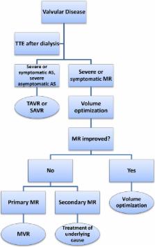
Recommendations for Management of Valvular Disease
Patients with clinically significant valvular abnormalities should be considered for
definitive management prior to transplantation. TAVR should be used as an alternative
to SAVR in patients at high or intermediate risk for surgery.117, 118 The decision
to proceed with TAVR or SAVR should be made in consultation with a cardiac surgeon
and an interventional cardiologist (Figure 3). Mitral valve surgery should be performed
only after documentation of severe valve dysfunction on echocardiography following
right heart catheterization showing normal filling pressures.
Figure 3
Algorithm for evaluation and treatment of valvular disease. AS indicates aortic stenosis;
MR, mitral regurgitation; MVR, mitral valve repair/replacement; SAVR, surgical aortic
valve replacement; TAVR, transcatheter aortic valve replacement; TTE, transthoracic
echocardiogram.
PH in Patients With ESRD
PH is common in patients with ESRD, and multiple studies have estimated the prevalence
to be 26% to 48% depending on the mean age of the population studied and the time
spent on dialysis.4, 119 The majority of PH observed in patients receiving RRT occurs
in patients with arteriovenous fistulae (AVF) for hemodialysis. Patients on peritoneal
dialysis also have a higher incidence of PH compared with the general population.4,
119 Several factors place patients with ESRD at risk for the development of PH: placement
of AVF, chronic hypervolemia, and anemia. These risk factors can lead to a state of
high cardiac output, which can further contribute to the development of PH. It is
essential to dialyze patients to their dry weights to prevent chronic volume overload
and reduce the risk of development of PH, which is frequently observed in this patient
population.120, 121 Compression of AVF for 1 minute has been shown to decrease cardiac
output and pulmonary arterial pressure and may be a useful diagnostic maneuver to
determine the reversibility of PH.122 Given the massive capacitance of the pulmonary
vasculature, increased cardiac output alone might not be the only driving force for
the development of PH in patients on dialysis.120 Endothelial dysfunction caused by
decreased nitric oxide production may also play a role122. It has been shown that
patients with PH on dialysis have reduced serum levels of nitric oxide both before
and after hemodialysis compared with patients on dialysis without PH.122 This suggests
that the uremic environment may reduce the capacitance of the pulmonary vasculature,
predisposing patients on dialysis with high cardiac outputs to the development of
PH.119, 122
Treatment of PH
Development of PH is associated with significant morbidity and mortality.121, 122,
123, 124, 125 Patients on dialysis with PH have significantly lower survival rates
than their counterparts without PH, with respective survival rates of 78.6% versus
96.5% at 1 year, 42.9% versus 78.8% at 3 years, and 25.5% versus 66.4% at 5 years.126
Thus, patients with ESRD and severe PH should be referred to a PH specialist for PH‐specific
therapies. Unfortunately, therapeutic options for patients with ESRD and PH are limited.
Treatments such as phosphodiesterase type 5 inhibitors or endothelin receptor antagonists
have not been studied specifically in patients with ESRD and PH. Surgical reduction
of AVF should be considered in patients with very high cardiac output in whom improvements
in cardiac output and PH by temporary AVF closure has been shown.121, 122, 126, 127
An AVF flow rate ≥2 L/min and cardiac output of ≥8 L/min place patients at high risk
of high‐output cardiac failure.128, 129 The definitive treatment for PH in this population
is renal transplantation if the etiology is secondary to high cardiac output from
AVF. These patients should be considered for renal transplantation as soon as possible.123,
124, 126
Evidence‐based guidelines for the perioperative management of patients with PH are
lacking because the AHA/ACC practice guidelines for noncardiac surgery do not list
PH as an independent risk factor for postoperative complications.125 Several small
studies, however, have suggested that PH is a risk factor for increased peri‐ and
postoperative morbidity and mortality. According to the AHA/ACC recommendations specifically
addressing cardiac disease evaluation among kidney transplantation candidates, right
heart catheterization is reasonable to pursue to confirm echocardiographic evidence
of elevated pulmonary arterial pressures.20 Right heart catheterization is also warranted
to assess the severity of PH before transplantation to determine whether there is
an association with a state of high cardiac output.20, 125 Consultation with a PH
specialist should also be considered early because therapy with phosphodiesterase
type 5 inhibitors or endothelin receptor antagonists may be needed to facilitate renal
transplantation in patients with refractory PH not secondary to AVF‐dependent high
cardiac output.121 During surgery, systemic hypotension or abrupt increases in pulmonary
artery pressures can cause right ventricular overload and lead to right ventricular
systolic dysfunction and decreased cardiac output. Therefore, intraoperative invasive
hemodynamic monitoring of pulmonary circulation should be considered.125 Nevertheless,
renal transplantation has been shown to be curative for PH under certain circumstances.
If the pulmonary pressures do not preclude a surgical procedure, renal transplantation
should be pursued aggressively to improve morbidity and mortality in this group of
patients.
Recommendations for Management of PH
Evidence of PH on echocardiogram (≥40 mm Hg) should be confirmed with repeat echocardiography
following hemodialysis to ensure that PH is not simply caused by volume overload (Figure 4).
If pulmonary artery pressures remain elevated despite optimization of volume status
by dialysis, right heart catheterization to assess severity and potential etiology
of PH should be performed. Severe PH (mean pulmonary artery pressure ≥40 mm Hg) in
the setting of elevated pulmonary capillary wedge pressure (≥18 mm Hg) should be treated
with more aggressive diuresis to optimize volume status, at times requiring inpatient
admission to perform daily dialysis. When PH is present in the absence of elevated
pulmonary capillary wedge pressure but with high cardiac output (>8 L/min), attention
should be paid to the AVF. Evidence of decreased cardiac output and improved pulmonary
pressures acutely during AVF occlusion in the catheterization laboratory are suggestive
of AVF as the etiology of PH, and surgical revision should be considered. Patients
with PH with normal left atrial pressures and normal cardiac output should undergo
reversibility testing with intravenous and with or without inhaled vasodilators to
determine the potential response to medical therapy. Patients with severe PH should
be referred to a PH specialist for help with perioperative management. A multidisciplinary
approach for perioperative management should be considered, including consultation
with anesthesiology to help determine the optimal intraoperative plan of care.125
Figure 4
Algorithm for evaluation and treatment of PH. AVF indicates arteriovenous fistula;
CO, cardiac output; ePASP, estimated pulmonary artery systolic pressure (echocardiography);
mPAP, mean pulmonary artery pressure; PCWP, pulmonary capillary wedge pressure; PH,
pulmonary hypertension; RHC, right heart catheterization; TTE, transthoracic echocardiogram.
Conclusions
Cardiovascular disease processes are highly prevalent and have major negative impacts
on clinical outcomes in patients with advanced CKD. Nevertheless, optimal cardiovascular
management in this population remains challenging due to the absence of data from
randomized clinical trials, from which this high‐risk group continues to be excluded.
Encouraging data on improvement of cardiovascular outcomes after successful renal
transplantation with appropriate cardiovascular workup and management highlights the
urgent need for clinical trials to investigate a wide array of unresolved clinical
issues related to cardiovascular pathologies in advanced CKD.
Disclosures
None.
Related collections
Most cited references94
- Record: found
- Abstract: found
- Article: not found
Evaluation and management of chronic kidney disease: synopsis of the kidney disease: improving global outcomes 2012 clinical practice guideline.
Paul E. Stevens (2013)
- Record: found
- Abstract: found
- Article: not found
Rosuvastatin and cardiovascular events in patients undergoing hemodialysis.
Bengt C Fellström, Alan Jardine, Roland E Schmieder … (2009)
- Record: found
- Abstract: not found
- Article: not found
Atherosclerosis is an inflammatory disease
Russell Ross (1999)