19
views
0
recommends
+1 Recommend
1 collections
    0
    shares

      International Journal of Nanomedicine (submit here)

      This international, peer-reviewed Open Access journal by Dove Medical Press focuses on the application of nanotechnology in diagnostics, therapeutics, and drug delivery systems throughout the biomedical field. Sign up for email alerts here.

      105,621 Monthly downloads/views I 7.033 Impact Factor I 10.9 CiteScore I 1.22 Source Normalized Impact per Paper (SNIP) I 1.032 Scimago Journal & Country Rank (SJR)

      • Record: found
      • Abstract: found
      • Article: found
      Is Open Access

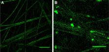
      Enhanced chondrocyte culture and growth on biologically inspired nanofibrous cell culture dishes

      research-article

      Read this article at

      ScienceOpenPublisherPMC
      Bookmark
          There is no author summary for this article yet. Authors can add summaries to their articles on ScienceOpen to make them more accessible to a non-specialist audience.

          Abstract

          Chondral and osteochondral defects affect a large number of people in which treatment options are currently limited. Due to its ability to mimic the natural nanofibrous structure of cartilage, this current in vitro study aimed at introducing a new scaffold, called XanoMatrix™, for cartilage regeneration. In addition, this same scaffold is introduced here as a new substrate onto which to study chondrocyte functions. Current studies on chondrocyte functions are limited due to nonbiologically inspired cell culture substrates. With its polyethylene terephthalate and cellulose acetate composition, good mechanical properties and nanofibrous structure resembling an extracellular matrix, XanoMatrix offers an ideal surface for chondrocyte growth and proliferation. This current study demonstrated that the XanoMatrix scaffolds promote chondrocyte growth and proliferation as compared with the Corning and Falcon surfaces normally used for chondrocyte cell culture. The XanoMatrix scaffolds also have greater hydrophobicity, three-dimensional surface area, and greater tensile strength, making them ideal candidates for alternative treatment options for chondral and osteochondral defects as well as cell culture substrates to study chondrocyte functions.

          Most cited references14

          • Record: found
          • Abstract: found
          • Article: not found

          A biomimetic three-dimensional woven composite scaffold for functional tissue engineering of cartilage.

          Tissue engineering seeks to repair or regenerate tissues through combinations of implanted cells, biomaterial scaffolds and biologically active molecules. The rapid restoration of tissue biomechanical function remains an important challenge, emphasizing the need to replicate structural and mechanical properties using novel scaffold designs. Here we present a microscale 3D weaving technique to generate anisotropic 3D woven structures as the basis for novel composite scaffolds that are consolidated with a chondrocyte-hydrogel mixture into cartilage tissue constructs. Composite scaffolds show mechanical properties of the same order of magnitude as values for native articular cartilage, as measured by compressive, tensile and shear testing. Moreover, our findings showed that porous composite scaffolds could be engineered with initial properties that reproduce the anisotropy, viscoelasticity and tension-compression nonlinearity of native articular cartilage. Such scaffolds uniquely combine the potential for load-bearing immediately after implantation in vivo with biological support for cell-based tissue regeneration without requiring cultivation in vitro.
            Bookmark
            • Record: found
            • Abstract: found
            • Article: not found

            Major biological obstacles for persistent cell-based regeneration of articular cartilage

            Hyaline articular cartilage, the load-bearing tissue of the joint, has very limited repair and regeneration capacities. The lack of efficient treatment modalities for large chondral defects has motivated attempts to engineer cartilage constructs in vitro by combining cells, scaffold materials and environmental factors, including growth factors, signaling molecules, and physical influences. Despite promising experimental approaches, however, none of the current cartilage repair strategies has generated long lasting hyaline cartilage replacement tissue that meets the functional demands placed upon this tissue in vivo. The reasons for this are diverse and can ultimately result in matrix degradation, differentiation or integration insufficiencies, or loss of the transplanted cells and tissues. This article aims to systematically review the different causes that lead to these impairments, including the lack of appropriate differentiation factors, hypertrophy, senescence, apoptosis, necrosis, inflammation, and mechanical stress. The current conceptual basis of the major biological obstacles for persistent cell-based regeneration of articular cartilage is discussed, as well as future trends to overcome these limitations.
              Bookmark
              • Record: found
              • Abstract: found
              • Article: not found

              Cytotoxicity of glutaraldehyde crosslinked collagen/poly(vinyl alcohol) films is by the mechanism of apoptosis.

              Collagen has been investigated as a potential natural biomaterial, because of its occurrence in the extracellular matrix. Collagen requires crosslinking in this context, by reagents that are often cytotoxic. Glutaraldehyde is one such agent that is potentially cytotoxic. The aim of this study was to determine the cause of poor cell attachment and growth on collagen/poly(vinyl alcohol) bioartificial composite films, when crosslinked with glutaraldehyde. Dehydrothermal crosslinking was used as a comparison. Human osteoblasts were observed to undergo apoptosis on glutaraldehyde crosslinked films dependent on concentration of collagen present. Higher collagen content resulted in higher levels of apoptosis with poor cell attachment and spreading of remaining cells. Post-treatment of films with 8% L-glutamic acid prevented the apoptotic response of osteoblasts and allowed attachment and spreading. The addition of 100 nM insulin-like growth factor-1 to the culture medium also prevented apoptosis. Glutaraldehyde toxicity of crosslinked collagen has been demonstrated in this study, the mechanism of which is apoptosis. This study indicates that poor biocompatibility and induction of apoptosis on collagen/poly(vinyl alcohol) films crosslinked by glutaraldehyde are attributed to glutaraldehyde components on the surface of the films (not residual glutaraldehyde), whose effects can be quenched by glutamic acid, and prevented by insulin-like growth factor-1. Copyright 2002 Wiley Periodicals, Inc.
                Bookmark

                Author and article information

                Journal
                Int J Nanomedicine
                Int J Nanomedicine
                International Journal of Nanomedicine
                International Journal of Nanomedicine
                Dove Medical Press
                1176-9114
                1178-2013
                2016
                04 February 2016
                : 11
                : 479-483
                Affiliations
                [1 ]Department of Chemical Engineering, Northeastern University, Boston, MA, USA
                [2 ]Center of Excellence for Advanced Materials Research, King Abdulaziz University, Jeddah, Saudi Arabia
                Author notes
                Correspondence: Thomas J Webster, Center of Excellence for Advanced Materials Research, King Abdulaziz University, Jeddah 215789, Saudi Arabia, Email th.webster@ 123456neu.edu
                Article
                ijn-11-479
                10.2147/IJN.S94000
                4751894
                26917958
                560f3578-ec58-4e64-b686-0bf08e910d24
                © 2016 Bhardwaj and Webster. This work is published and licensed by Dove Medical Press Limited

                The full terms of this license are available at https://www.dovepress.com/terms.php and incorporate the Creative Commons Attribution – Non Commercial (unported, v3.0) License ( http://creativecommons.org/licenses/by-nc/3.0/). By accessing the work you hereby accept the Terms. Non-commercial uses of the work are permitted without any further permission from Dove Medical Press Limited, provided the work is properly attributed.

                History
                Categories
                Original Research

                Molecular medicine
                chondrocytes,xanomatrix™,cell culture,substrates,biomimetic scaffolds
                Molecular medicine
                chondrocytes, xanomatrix™, cell culture, substrates, biomimetic scaffolds

                Comments

                Comment on this article