24
views
0
recommends
+1 Recommend
0 collections
    0
    shares
      • Record: found
      • Abstract: found
      • Article: found
      Is Open Access

      Anatomic Variants Mimicking Pathology on Echocardiography: Differential Diagnosis

      review-article
      , MD, , MD
      Journal of Cardiovascular Ultrasound
      Korean Society of Echocardiography
      Anatomic variation, Echocardiography

      Read this article at

      Bookmark
          There is no author summary for this article yet. Authors can add summaries to their articles on ScienceOpen to make them more accessible to a non-specialist audience.

          Abstract

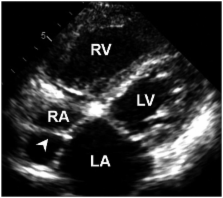
          Differentiation of normal from abnormal findings is critical in echocardiography. Anatomic variants occurring in normal cardiac developments often simulate pathologic entities. This review focuses on the differential diagnosis of normal anatomic structures from pathologic ones in echocardiography.

          Related collections

          Most cited references43

          • Record: found
          • Abstract: found
          • Article: not found

          Cardiac papillary fibroelastoma: a comprehensive analysis of 725 cases.

          With the advent of echocardiography, cardiac papillary fibroelastoma (CPF) is being increasingly reported. The demographics, clinical characteristics, pathological features, treatment, and prognosis of CPF are examined. Cases, case series and related articles on the subject in all languages were identified through a comprehensive literature search. Seven hundred twenty-five cases of CPF were identified. Males comprised 55% of patients. Highest prevalence was in the 8th decade of life. The valvular surface was the predominant locations of tumor. The most commonly involved valve was the aortic valve, followed by the mitral valve. The left ventricle was the predominant nonvalvular site involved. No clear risk factor for development of CPF has been reported. Size of the tumor varied from 2 mm to 70 mm. Clinically, CPFs have presented with transient ischemic attack, stroke, myocardial infarction, sudden death, heart failure, presyncope, syncope, pulmonary embolism, blindness, and peripheral embolism. Tumor mobility was the only independent predictor of CPF-related death or nonfatal embolization. Symptomatic patients should be treated surgically because the successful complete resection of CPF is curative and the long-term postoperative prognosis is excellent. The symptomatic patients who are not surgical candidates could be offered long-term oral anticoagulation, although no randomized controlled data are available on its efficacy. Asymptomatic patients could be treated surgically if the tumor is mobile, as the tumor mobility is the independent predictor of death or nonfatal embolization. Asymptomatic patients with nonmobile CPF could be followed-up closely with periodic clinical evaluation and echocardiography, and receive surgical intervention when symptoms develop or the tumor becomes mobile.
            Bookmark
            • Record: found
            • Abstract: found
            • Article: not found

            Papillary fibroelastoma: echocardiographic characteristics for diagnosis and pathologic correlation.

            We sought to determine the clinical and echocardiographic characteristics of papillary fibroelastoma (PFE). PFE is a rarely encountered cardiac tumor about which relatively little is known. Institutional records were reviewed for the years 1980 to 1995 for patients with pathologic or echocardiographic diagnosis of PFE. Group 1 included 17 patients with the pathologic diagnosis of PFE who also underwent echocardiography. Echocardiographic features of PFE were established in group 1. Group 2 included 37 patients with only echocardiographic evidence of PFE. In group 1, 7 (41.2%) of 17 patients had symptoms related to PFE. Neurologic events occurred in 5 (29.4%) of 17 patients. All patients had the tumor surgically removed. During follow-up, no new embolic events occurred. Echocardiographic characteristics of PFE included a small tumor (12.1 +/- 6.5 x 9.0 +/- 4.3 mm), usually pedunculated (14 [94%] of 17 patients) and mobile, with a homogeneous speckled pattern and a characteristic stippling along the edges. PFEs were most common on valvular surfaces (12 [60%] of 20 PFEs) but were not uncommon on other endocardial surfaces (8 [40%] of 20 PFEs). The tumor did not cause valvular dysfunction. In group 2, 16 (43%) of 37 patients were asymptomatic. Five patients (13.5%) had a previous neurologic event. During follow-up (mean 31 months, range 1 to 77), nine neurologic events occurred. PFEs are associated with embolism, can be diagnosed with echocardiography, are often an incidental clinical finding and do not cause valvular dysfunction.
              Bookmark
              • Record: found
              • Abstract: found
              • Article: not found

              Lipomatous hypertrophy of the interatrial septum: a prospective study of incidence, imaging findings, and clinical symptoms.

              Lipomatous hypertrophy of the interatrial septum (LHIS) is a benign disorder characterized by fat accumulation in the interatrial septum. It typically occurs in elderly, obese patients and may cause arrhythmia. The purpose of this study was to determine the imaging features of this cardiac pathology using multislice CT (MSCT), and its relationship with clinical findings. A total of 1,292 consecutive patients who underwent MSCT of the thorax from September 2001 to August 2002 were prospectively studied. Beside the analysis of patient records, the amount of fat in the interatrial septum, and its size and shape were determined. Among the 1,292 patients investigated, we found 28 (2.2%) with LHIS. The mean age of affected patients was 72.2 years. MSCT studies revealed a mass of fat attenuation with sharp margins and sparing of the fossa ovalis, resulting in a dumbbell shape in all patients. The median thickness of the interatrial septum was 32 mm (range, 20 to 62 mm), and the median craniocaudal extend was 62 mm (range, 51 to 89 mm). Twenty-one patients (75.0%) showed increased epicardial fat, 18 patients (64.3%) had significant pulmonary emphysema, and 13 of 21 patients (61.9%) showed ECG abnormalities. Three patients underwent functional cardiac MRI studies. In one patient, hemodynamic obstruction by LHIS was shown. MSCT scanning is a useful method to diagnose LHIS, a cardiac condition that in our series reached a 2.2% incidence. The lesion shows characteristic features, thus differentiating it from other cardiac tumors. Advanced age, obesity, pulmonary emphysema, and atrial arrhythmias are common additional findings in patients with LHIS.
                Bookmark

                Author and article information

                Journal
                J Cardiovasc Ultrasound
                J Cardiovasc Ultrasound
                JCU
                Journal of Cardiovascular Ultrasound
                Korean Society of Echocardiography
                1975-4612
                2005-9655
                September 2013
                30 September 2013
                : 21
                : 3
                : 103-112
                Affiliations
                Division of Cardiology, Department of Internal Medicine, College of Medicine, The Catholic University of Korea, Seoul, Korea.
                Author notes
                Address for Correspondence: Hae Ok Jung, Division of Cardiology, Department of Internal Medicine, College of Medicine, The Catholic University of Korea, 222 Banpo-daero, Seocho-gu, Seoul 137-701, Korea. Tel: +82-2-2258-6033, Fax: +82-2-591-1506, hojheart@ 123456catholic.ac.kr
                Article
                10.4250/jcu.2013.21.3.103
                3816159
                24198915
                56d4a5ab-bf1b-49f0-8885-4b70c4b783a6
                Copyright © 2013 Korean Society of Echocardiography

                This is an Open Access article distributed under the terms of the Creative Commons Attribution Non-Commercial License ( http://creativecommons.org/licenses/by-nc/3.0/) which permits unrestricted non-commercial use, distribution, and reproduction in any medium, provided the original work is properly cited.

                History
                : 23 August 2013
                : 17 September 2013
                : 17 September 2013
                Categories
                Review

                Cardiovascular Medicine
                anatomic variation,echocardiography
                Cardiovascular Medicine
                anatomic variation, echocardiography

                Comments

                Comment on this article