2
views
0
recommends
+1 Recommend
0 collections
    0
    shares
      • Record: found
      • Abstract: found
      • Article: found
      Is Open Access

      Monocyte to high-density lipoprotein and neutrophil-to-lymphocyte ratios in patients with acute central serous chorioretinopathy

      research-article

      Read this article at

      Bookmark
          There is no author summary for this article yet. Authors can add summaries to their articles on ScienceOpen to make them more accessible to a non-specialist audience.

          Abstract

          Purpose:

          To investigate monocyte to high-density lipoprotein (HDL) ratio (MHR) and neutrophil-to-lymphocyte ratio (NLR) as indicators of systemic inflammation in acute central serous chorioretinopathy (CSC).

          Methods:

          The HDL levels, hematological profiles, erythrocyte sedimentation rates (ESR), and C-reactive protein (CRP) levels of 38 patients with acute CSC (Group I) and 38 controls without CSC (Group II) were measured.

          Results:

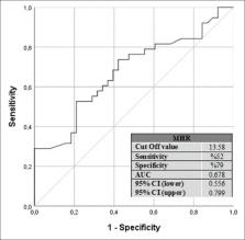
          MHRs were significantly higher in Group I (13.30 ± 2.95) than in Group II (11.52 ± 2.42, P = 0.005), whereas NLRs, CRP values, and ESR values did not significantly differ between the groups ( P = 0.726, P = 0.219, and P = 0.441, respectively). Multivariate analysis revealed that the MHR was an independent predictor of acute CSC (OR = 1.266, 95% CI = 1.054-1.521, P = 0.012).

          Conclusion:

          Indicating an association between increased MHRs and acute CSC, the MHR might represent simple, inexpensive, reliable biomarkers of inflammation in acute CSC.

          Related collections

          Most cited references36

          • Record: found
          • Abstract: found
          • Article: not found

          Central serous chorioretinopathy: Recent findings and new physiopathology hypothesis.

          Central serous chorioretinopathy (CSCR) is a major cause of vision threat among middle-aged male individuals. Multimodal imaging led to the description of a wide range of CSCR manifestations, and highlighted the contribution of the choroid and pigment epithelium in CSCR pathogenesis. However, the exact molecular mechanisms of CSCR have remained uncertain. The aim of this review is to recapitulate the clinical understanding of CSCR, with an emphasis on the most recent findings on epidemiology, risk factors, clinical and imaging diagnosis, and treatments options. It also gives an overview of the novel mineralocorticoid pathway hypothesis, from animal data to clinical evidences of the biological efficacy of oral mineralocorticoid antagonists in acute and chronic CSCR patients. In rodents, activation of the mineralocorticoid pathway in ocular cells either by intravitreous injection of its specific ligand, aldosterone, or by over-expression of the receptor specifically in the vascular endothelium, induced ocular phenotypes carrying many features of acute CSCR. Molecular mechanisms include expression of the calcium-dependent potassium channel (KCa2.3) in the endothelium of choroidal vessels, inducing subsequent vasodilation. Inappropriate or over-activation of the mineralocorticoid receptor in ocular cells and other tissues (such as brain, vessels) could link CSCR with the known co-morbidities observed in CSCR patients, including hypertension, coronary disease and psychological stress.
            Bookmark
            • Record: found
            • Abstract: found
            • Article: not found

            Central serous chorioretinopathy.

            Central serous chorioretinopathy (CSC) is a disease of the retina characterized by serous detachment of the neurosensory retina secondary to one or more focal lesions of the retinal pigment epithelium (RPE). CSC occurs most frequently in mid-life and more often in men than in women. Major symptoms are blurred vision, usually in one eye only and perceived typically by the patient as a dark spot in the centre of the visual field with associated micropsia and metamorphopsia. Normal vision often recurs spontaneously within a few months. The condition can be precipitated by psychosocial stress and hypercortisolism. Ophthalmoscopic signs of CSC range from mono- or paucifocal RPE lesions with prominent elevation of the neurosensory retina by clear fluid - typical of cases of recent onset - to shallow detachments overlying large patches of irregularly depigmented RPE. The spectrum of lesions includes RPE detachments. Granular or fibrinous material may accumulate in the subretinal cavity. Serous detachment often resolves spontaneously. From first contact, counselling about the potential relation to stress and glucocorticoid medication is warranted. After 3 months without resolution of acute CSC or in chronic CSC, treatment should be considered. Resolution of detachment can usually be achieved in acute CSC by focal photocoagulation of leaking RPE lesions or, in chronic CSC, by photodynamic therapy. The effect of therapy on long-term visual outcome is insufficiently documented. Reattachment within 4 months of onset is considered a relevant therapeutic target because prolonged detachment is associated with photoreceptor atrophy. This suggests that the value of treatment depends upon proper selection of cases that will not resolve without therapy. Chronic CSC may be difficult to differentiate from occult choroidal neovascularization secondary to CSC. Patients with chronic CSC who receive glucocorticoid treatment for systemic disease can often be managed without having to discontinue this medication.
              Bookmark
              • Record: found
              • Abstract: found
              • Article: not found

              Digital indocyanine green videoangiography of central serous chorioretinopathy.

              The pathogenesis of central serous chorioretinopathy (CSC) is poorly understood. Abnormalities in the choroidal circulation have been hypothesized to be causative factors. Fluorescein angiography has not been particularly useful in identifying specific choroidal defects in CSC, largely because of inherent limitations in imaging with this technique. Recent technologic advances in digital indocyanine green videoangiography allow enhanced imaging of the choroid and other subretinal structures in comparison with fluorescein angiography. We performed digital indocyanine green videoangiography in 29 consecutive eyes with CSC and compared our results with clinical and fluorescein angiographic findings. Several newly recognized subretinal abnormalities in CSC were noted with digital indocyanine green videoangiography, including (1) presumed hyperpermeability of the choroidal circulation surrounding active retinal pigment epithelial leaks, (2) additional focal and multifocal areas of presumed choroidal hyperpermeability not associated with abnormalities detectable by fluorescein angiography or clinical examination, and (3) multiple presumed "occult" serous retinal pigment epithelial detachments with a characteristic indocyanine green videoangiographic pattern. We suggest that the pathogenesis of CSC may be due to a choroidal vascular hyperpermeability with and without associated active pigment epithelial leaks and multiple presumed "occult" serous retinal pigment epithelial detachments. Based on these findings, a hypothetical model can be constructed related to the pathogenesis of CSC, beginning with choroidal abnormalities that secondarily affect the retinal pigment epithelium and neurosensory retina.
                Bookmark

                Author and article information

                Journal
                Indian J Ophthalmol
                Indian J Ophthalmol
                IJO
                Indian Journal of Ophthalmology
                Wolters Kluwer - Medknow (India )
                0301-4738
                1998-3689
                May 2020
                20 April 2020
                : 68
                : 5
                : 854-858
                Affiliations
                [1]Department of Ophthalmology, The Kayseri City Hospital, Kayseri, Turkey
                Author notes
                Correspondence to: Dr. Ender Sirakaya, Department of Ophthalmology, The Kayseri City Hospital, Kayseri, Turkey. E-mail: esirakaya@ 123456gmail.com
                Article
                IJO-68-854
                10.4103/ijo.IJO_1327_19
                7350498
                32317461
                56d5393d-1d84-4ed6-a6d4-a8edb85f3b02
                Copyright: © 2020 Indian Journal of Ophthalmology

                This is an open access journal, and articles are distributed under the terms of the Creative Commons Attribution-NonCommercial-ShareAlike 4.0 License, which allows others to remix, tweak, and build upon the work non-commercially, as long as appropriate credit is given and the new creations are licensed under the identical terms.

                History
                : 20 July 2019
                : 16 September 2019
                : 01 November 2019
                Categories
                Original Article

                Ophthalmology & Optometry
                acute central serous chorioretinopathy,monocyte to high-density lipoprotein ratio,neutrophil-to-lymphocyte ratio,systemic inflammation

                Comments

                Comment on this article