24
views
0
recommends
+1 Recommend
0 collections
    0
    shares
      • Record: found
      • Abstract: found
      • Article: found
      Is Open Access

      Treatment of glaucomatous optic nerve damage using ginsenoside Rg1 mediated by ultrasound targeted microbubble destruction

      research-article

      Read this article at

      Bookmark
          There is no author summary for this article yet. Authors can add summaries to their articles on ScienceOpen to make them more accessible to a non-specialist audience.

          Abstract

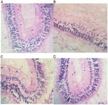
          The treatment of glaucomatous optic nervedamage using ginsenoside Rg1 mediated by ultrasound targeted microbubbles destruction was evaluated. Thirty healthy New Zealand white rabbits were subjected to injection of 0.3% carbomer solution to establish glaucomatous optic nerve damage model. Rabbits were divided into 5 groups: control group, model group, model group + intravitreal injection of nerve growth factor (NGF) group, model group + intravitreal injection of ginsenoside Rg1 group (Rg1 group), model group + intravitreal injection of ginsenoside Rg1 + ultrasound microbubble group (ultrasound group), model group + ultrasound targeted microbubble destruction (ultrasound group). Intraocular pressures were compared at 1, 2 and 4 weeks after model establishment. Rabbits were sacrificed 4 weeks after model establishment to collect retinal tissue for H&E staining. Histological changes were observed and the retinal thickness was measured. Contents of malondialdehyde (MDA), superoxide dismutase (SOD) and nitric oxide (NO) were measured by ELISA. Intraocular pressure was significantly higher in model group than in control group at 1 week (P<0.05). Intraocular pressure was significantly lower in the ultrasound group than in NGF group and Rg1 group at all time-points (P<0.05). The number of ganglion cells in model group was decreased significantly. Number of nuclear layer cells was significantly reduced. Thickest retina was found in control group and model group was the thinnest (P<0.05). Contents of MDA and NO in model group were significantly higher than those in NCF group and Rg1 group. SOD content in control group was higher than that in ultrasound group and model group (P<0.05). In conclusion, treatment of glaucomatous optic nerve damage using ginsenoside Rg1 mediated by ultrasound targeted microbubble destruction can reduce the level of oxidative stress, relieve intraocular pressure and reduce ganglion cell damage.

          Related collections

          Most cited references18

          • Record: found
          • Abstract: found
          • Article: found
          Is Open Access

          Neuroprotection, Growth Factors and BDNF-TrkB Signalling in Retinal Degeneration

          Neurotrophic factors play key roles in the development and survival of neurons. The potent neuroprotective effects of neurotrophic factors, including brain-derived neurotrophic factor (BDNF), ciliary neurotrophic factor (CNTF), glial cell-line derived neurotrophic factor (GDNF) and nerve growth factor (NGF), suggest that they are good therapeutic candidates for neurodegenerative diseases. Glaucoma is a neurodegenerative disease of the eye that causes irreversible blindness. It is characterized by damage to the optic nerve, usually due to high intraocular pressure (IOP), and progressive degeneration of retinal neurons called retinal ganglion cells (RGCs). Current therapy for glaucoma focuses on reduction of IOP, but neuroprotection may also be beneficial. BDNF is a powerful neuroprotective agent especially for RGCs. Exogenous application of BDNF to the retina and increased BDNF expression in retinal neurons using viral vector systems are both effective in protecting RGCs from damage. Furthermore, induction of BDNF expression by agents such as valproic acid has also been beneficial in promoting RGC survival. In this review, we discuss the therapeutic potential of neurotrophic factors in retinal diseases and focus on the differential roles of glial and neuronal TrkB in neuroprotection. We also discuss the role of neurotrophic factors in neuroregeneration.
            Bookmark
            • Record: found
            • Abstract: found
            • Article: not found

            Expression and signaling of NGF in the healthy and injured retina.

            This review summarizes the present knowledge concerning the retinal localization of the nerve growth factor (NGF), its precursor proNGF, and the receptors TrkA and p75NTR in the developing and mature rodent retina. We further discuss the changes in the expression of NGF and the receptors in experimental models of retinal disorders and diseases like inherited retinitis pigmentosa, retinal detachment, glaucoma, and diabetic retinopathy. Since proNGF is now recognized as a bioactive signaling molecule which induces cell death through p75NTR activation, the role of proNGF in the induction of retinal cell loss under neurodegenerative conditions is also highlighted. In addition, we present the evidences for a potential therapeutic intervention with NGF for the treatment of retinal neurodegenerative diseases. Different strategies have been developed and experimentally tested in mice and rats in order to reduce cell loss and Müller cell gliosis, e.g., increasing the availability of endogenous NGF, administration of exogenous NGF, activation of TrkA, and inhibition of p75NTR. Here, we discuss the several lines of evidence supporting a protective effect of NGF on retinal cell loss, with specific emphasis on photoreceptor and retinal ganglion cell degeneration. A better understanding of the mechanisms underlying the effects of NGF and proNGF in the modulation of neurodegeneration and gliosis in the retina will help to develop efficient therapeutic strategies for various retinal diseases.
              Bookmark
              • Record: found
              • Abstract: found
              • Article: found
              Is Open Access

              Exploring Serum Levels of Brain Derived Neurotrophic Factor and Nerve Growth Factor Across Glaucoma Stages

              Purpose To investigate the serum levels of Brain Derived Neurotrophic Factor (BDNF) and Nerve Growth Factor (NGF) in patients affected by primary open angle glaucoma with a wide spectrum of disease severity compared to healthy controls and to explore their relationship with morphological and functional glaucoma parameters. Materials and Methods 45 patients affected by glaucoma at different stages and 15 age-matched healthy control subjects underwent visual field testing, peripapillary retinal nerve fibre layer thickness measurement using Spectral Domain Optical Coherence Tomography and blood collection for both neurotrophins detection by Enzyme-Linked Immunosorbent Assay. Statistical analysis and association between biostrumental and biochemical data were investigated. Results Serum levels of BDNF in glaucoma patients were significantly lower than those measured in healthy controls (261.2±75.0 pg/ml vs 313.6±79.6 pg/ml, p = 0.03). Subgroups analysis showed that serum levels of BDNF were significantly lower in early (253.8±40.7 pg/ml, p = 0.019) and moderate glaucoma (231.3±54.3 pg/ml, p = 0.04) but not in advanced glaucoma (296.2±103.1 pg/ml, p = 0.06) compared to healthy controls. Serum levels of NGF in glaucoma patients were significantly lower than those measured in the healthy controls (4.1±1 pg/mL vs 5.5±1.2 pg/mL, p = 0.01). Subgroups analysis showed that serum levels of NGF were significantly lower in early (3.5±0.9 pg/mL, p = 0.0008) and moderate glaucoma (3.8±0.7 pg/ml, p<0.0001) but not in advanced glaucoma (5.0±0.7 pg/ml, p = 0.32) compared to healthy controls. BDNF serum levels were not related to age, visual field mean deviation or retinal nerve fibre layer thickness either in glaucoma or in controls while NGF levels were significantly related to visual field mean deviation in the glaucoma group (r2 = 0.26, p = 0.004). Conclusions BDNF and NGF serum levels are reduced in the early and moderate glaucoma stages, suggesting the possibility that both factors could be further investigated as potential circulating biomarkers for the early detection of glaucoma.
                Bookmark

                Author and article information

                Journal
                Exp Ther Med
                Exp Ther Med
                ETM
                Experimental and Therapeutic Medicine
                D.A. Spandidos
                1792-0981
                1792-1015
                January 2018
                27 October 2017
                27 October 2017
                : 15
                : 1
                : 300-304
                Affiliations
                Section One, Department of Ophthalmology, Cangzhou Central Hospital, Cangzhou, Hebei 061001, P.R. China
                Author notes
                Correspondence to: Dr Lianfeng Wang, Section One, Department of Ophthalmology, Cangzhou Central Hospital, 16 Xinhuaxi Road, Cangzhou, Hebei 061001, P.R. China, E-mail: wma2sq@ 123456163.com
                Article
                ETM-0-0-5386
                10.3892/etm.2017.5386
                5763670
                29375689
                56dd38e2-febb-4690-93e1-7fd291631b6d
                Copyright: © Wang et al.

                This is an open access article distributed under the terms of the Creative Commons Attribution-NonCommercial-NoDerivs License, which permits use and distribution in any medium, provided the original work is properly cited, the use is non-commercial and no modifications or adaptations are made.

                History
                : 21 June 2017
                : 10 October 2017
                Categories
                Articles

                Medicine
                ultrasound targeted microbubble destruction,ginsenoside rg1,nerve growth factor,glaucoma,malondialdehyde,superoxide dismutase,nitric oxide

                Comments

                Comment on this article