15
views
0
recommends
+1 Recommend
0 collections
    0
    shares
      • Record: found
      • Abstract: found
      • Article: found
      Is Open Access

      Mitophagy-mediated adipose inflammation contributes to type 2 diabetes with hepatic insulin resistance

      research-article

      Read this article at

      Bookmark
          There is no author summary for this article yet. Authors can add summaries to their articles on ScienceOpen to make them more accessible to a non-specialist audience.

          Abstract

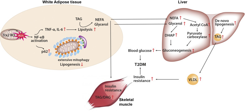
          Adipose-specific deletion of mitochondrial redox Trx2 induces excessive mitophagy in white adipose tissue with increased inflammation and increased lipolysis, promoting hepatic glucose production and development of T2DM with hepatic steatosis. Administration of NF-κB inhibitor prevents adipose mitophagy and ameliorates T2DM progression.

          Abstract

          White adipose tissues (WAT) play crucial roles in maintaining whole-body energy homeostasis, and their dysfunction can contribute to hepatic insulin resistance and type 2 diabetes mellitus (T2DM). However, the mechanisms underlying these alterations remain unknown. By analyzing the transcriptome landscape in human adipocytes based on available RNA-seq datasets from lean, obese, and T2DM patients, we reveal elevated mitochondrial reactive oxygen species (ROS) pathway and NF-κB signaling with altered fatty acid metabolism in T2DM adipocytes. Mice with adipose-specific deletion of mitochondrial redox Trx2 develop hyperglycemia, hepatic insulin resistance, and hepatic steatosis. Trx2-deficient WAT exhibited excessive mitophagy, increased inflammation, and lipolysis. Mechanistically, mitophagy was induced through increasing ROS generation and NF-κB–dependent accumulation of autophagy receptor p62/SQSTM1, which recruits damaged mitochondria with polyubiquitin chains. Importantly, administration of ROS scavenger or NF-κB inhibitor ameliorates glucose and lipid metabolic disorders and T2DM progression in mice. Taken together, this study reveals a previously unrecognized mechanism linking mitophagy-mediated adipose inflammation to T2DM with hepatic insulin resistance.

          Graphical Abstract

          Related collections

          Most cited references69

          • Record: found
          • Abstract: found
          • Article: not found

          Mechanisms of Insulin Action and Insulin Resistance

          The 1921 discovery of insulin was a Big Bang from which a vast and expanding universe of research into insulin action and resistance has issued. In the intervening century, some discoveries have matured, coalescing into solid and fertile ground for clinical application; others remain incompletely investigated and scientifically controversial. Here, we attempt to synthesize this work to guide further mechanistic investigation and to inform the development of novel therapies for type 2 diabetes (T2D). The rational development of such therapies necessitates detailed knowledge of one of the key pathophysiological processes involved in T2D: insulin resistance. Understanding insulin resistance, in turn, requires knowledge of normal insulin action. In this review, both the physiology of insulin action and the pathophysiology of insulin resistance are described, focusing on three key insulin target tissues: skeletal muscle, liver, and white adipose tissue. We aim to develop an integrated physiological perspective, placing the intricate signaling effectors that carry out the cell-autonomous response to insulin in the context of the tissue-specific functions that generate the coordinated organismal response. First, in section II, the effectors and effects of direct, cell-autonomous insulin action in muscle, liver, and white adipose tissue are reviewed, beginning at the insulin receptor and working downstream. Section III considers the critical and underappreciated role of tissue crosstalk in whole body insulin action, especially the essential interaction between adipose lipolysis and hepatic gluconeogenesis. The pathophysiology of insulin resistance is then described in section IV. Special attention is given to which signaling pathways and functions become insulin resistant in the setting of chronic overnutrition, and an alternative explanation for the phenomenon of ‟selective hepatic insulin resistanceˮ is presented. Sections V, VI, and VII critically examine the evidence for and against several putative mediators of insulin resistance. Section V reviews work linking the bioactive lipids diacylglycerol, ceramide, and acylcarnitine to insulin resistance; section VI considers the impact of nutrient stresses in the endoplasmic reticulum and mitochondria on insulin resistance; and section VII discusses non-cell autonomous factors proposed to induce insulin resistance, including inflammatory mediators, branched-chain amino acids, adipokines, and hepatokines. Finally, in section VIII, we propose an integrated model of insulin resistance that links these mediators to final common pathways of metabolite-driven gluconeogenesis and ectopic lipid accumulation.
            Bookmark
            • Record: found
            • Abstract: found
            • Article: not found

            Crosstalk of reactive oxygen species and NF-κB signaling.

            NF-κB proteins are a family of transcription factors that are of central importance in inflammation and immunity. NF-κB also plays important roles in other processes, including development, cell growth and survival, and proliferation, and is involved in many pathological conditions. Reactive Oxygen Species (ROS) are created by a variety of cellular processes as part of cellular signaling events. While certain NF-κB-regulated genes play a major role in regulating the amount of ROS in the cell, ROS have various inhibitory or stimulatory roles in NF-κB signaling. Here we review the regulation of ROS levels by NF-κB targets and various ways in which ROS have been proposed to impact NF-κB signaling pathways.
              Bookmark
              • Record: found
              • Abstract: found
              • Article: not found

              Inflammation, metaflammation and immunometabolic disorders

              Proper regulation and management of energy, substrate diversity and quantity, as well as macromolecular synthesis and breakdown processes, are fundamental to cellular and organismal survival and are paramount to health. Cellular and multicellular organization are defended by the immune response, a robust and critical system
                Bookmark

                Author and article information

                Contributors
                Role: ConceptualizationRole: Data curationRole: Formal analysisRole: InvestigationRole: MethodologyRole: Project administrationRole: SupervisionRole: ValidationRole: VisualizationRole: Writing - original draftRole: Writing - review & editing
                Role: ConceptualizationRole: Formal analysisRole: InvestigationRole: ValidationRole: VisualizationRole: Writing - original draft
                Role: ConceptualizationRole: Data curationRole: Formal analysisRole: InvestigationRole: Project administrationRole: SupervisionRole: ValidationRole: Visualization
                Role: ConceptualizationRole: Data curationRole: Formal analysisRole: InvestigationRole: MethodologyRole: Project administrationRole: ResourcesRole: Software
                Role: InvestigationRole: Resources
                Role: InvestigationRole: Writing - review & editing
                Role: ConceptualizationRole: Funding acquisitionRole: InvestigationRole: MethodologyRole: Project administrationRole: ResourcesRole: SupervisionRole: ValidationRole: VisualizationRole: Writing - original draftRole: Writing - review & editing
                Role: ConceptualizationRole: Data curationRole: Formal analysisRole: Funding acquisitionRole: InvestigationRole: MethodologyRole: Project administrationRole: ResourcesRole: SoftwareRole: SupervisionRole: ValidationRole: VisualizationRole: Writing - original draftRole: Writing - review & editing
                Journal
                J Exp Med
                J Exp Med
                jem
                The Journal of Experimental Medicine
                Rockefeller University Press
                0022-1007
                1540-9538
                01 March 2021
                14 December 2020
                : 218
                : 3
                : e20201416
                Affiliations
                [1 ]Department of Pathology, Yale School of Medicine, New Haven, CT
                [2 ]Department of Cellular and Molecular Physiology, Yale School of Medicine, New Haven, CT
                [3 ]Department of Internal Medicine, Yale School of Medicine, New Haven, CT
                Author notes
                Correspondence to Wang Min: mike.wang388@ 123456gmail.com
                Gerald I. Shulman: gerald.shulman@yale.edu

                Disclosures: G. Shulman reported grants from NIH (R01 DK119668, R01 DK116774, R01 DK113984, R01 DK114793, RC2 DK120534, and P30 DK045735), Gilead Sciences, Inc., AstraZeneca, and Merck & Co., Inc.; non-financial support from Gilead Sciences, Inc., AstraZeneca, Merck & Co., Inc., Janssen Research & Development, and Novo Nordisk; and personal fees from Gilead Sciences, Inc., AstraZeneca, Merck & Co., Inc., Janssen Research & Development, Novo Nordisk, iMetabolic Biopharma Corp., Maze Therapeutics, Inc., Generian Pharmaceuticals, Ionis Pharmaceuticals, Inc., BridgeBio, Esperion, 89Bio, Inc., Nimbus Discovery, Inc., Staten Biotechnology, Longitude Capital, Celgene Corp., and Aegerion Pharmaceuticals during the conduct of the study. No other disclosures were reported.

                [*]

                F. He, Y. Huang, Z. Song, and H.J. Zhou contributed equally to this paper.

                Author information
                https://orcid.org/0000-0003-3207-1295
                https://orcid.org/0000-0001-5147-1302
                https://orcid.org/0000-0003-3742-0570
                https://orcid.org/0000-0003-3071-9473
                https://orcid.org/0000-0001-9141-7458
                https://orcid.org/0000-0003-0748-8064
                https://orcid.org/0000-0003-1529-5668
                https://orcid.org/0000-0002-2479-6096
                Article
                jem.20201416
                10.1084/jem.20201416
                7927432
                33315085
                576f0697-1b3e-46c1-bc82-f0a1c05e602f
                © 2020 He et al.

                This article is distributed under the terms of an Attribution–Noncommercial–Share Alike–No Mirror Sites license for the first six months after the publication date (see http://www.rupress.org/terms/). After six months it is available under a Creative Commons License (Attribution–Noncommercial–Share Alike 4.0 International license, as described at https://creativecommons.org/licenses/by-nc-sa/4.0/).

                History
                : 06 July 2020
                : 02 September 2020
                : 19 October 2020
                Page count
                Pages: 22
                Funding
                Funded by: National Institutes of Health, DOI http://dx.doi.org/10.13039/100000002;
                Award ID: R01 HL109420
                Award ID: HL115148
                Award ID: R01 DK113984
                Award ID: R01 DK114793
                Award ID: R01 DK116774
                Award ID: P30 DK045735
                Funded by: American Heart Association, DOI http://dx.doi.org/10.13039/100000968;
                Award ID: 19CDA34760284
                Categories
                Article
                Metabolism

                Medicine
                Medicine

                Comments

                Comment on this article