26
views
0
recommends
+1 Recommend
0 collections
    0
    shares
      • Record: found
      • Abstract: found
      • Article: found
      Is Open Access

      Comparative analysis of minimal residual disease detection by multiparameter flow cytometry and enhanced ASO RQ-PCR in multiple myeloma

      research-article

      Read this article at

      Bookmark
          There is no author summary for this article yet. Authors can add summaries to their articles on ScienceOpen to make them more accessible to a non-specialist audience.

          Abstract

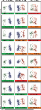
          Multiparameter flow cytometry (MFC) and allele-specific oligonucleotide real-time quantitative PCR (ASO RQ-PCR) are the two most sensitive methods to detect minimal residual disease (MRD) in multiple myeloma (MM). We compared these methods in 129 paired post-therapy samples from 22 unselected, consecutive MM patients in complete/near complete remission. Appropriate immunophenotypic and ASO RQ-PCR-MRD targets could be detected and MRD analyses constructed for all patients. The high PCR coverage could be achieved by gradual widening of the primer sets used for clonality detection. In addition, for 13 (55%) of the patients, reverse orientation of the ASO primer and individual design of the TaqMan probe improved the sensitivity and specificity of ASO RQ-PCR analysis. A significant nonlinear correlation prevailed between MFC-MRD and PCR-MRD when both were positive. Discordance between the methods was found in 32 (35%) paired samples, which were negative by MFC-MRD, but positive by ASO RQ-PCR. The findings suggest that with the described technique, ASO RQ-PCR can be constructed for all patients with MM. ASO RQ-PCR is slightly more sensitive in MRD detection than 6−10-color flow cytometry. Owing to technical demands ASO RQ-PCR could be reserved for patients in immunophenotypic remission, especially in efficacy comparisons between different drugs and treatment modalities.

          Related collections

          Most cited references18

          • Record: found
          • Abstract: found
          • Article: found
          Is Open Access

          EuroFlow antibody panels for standardized n-dimensional flow cytometric immunophenotyping of normal, reactive and malignant leukocytes

          Most consensus leukemia & lymphoma antibody panels consist of lists of markers based on expert opinions, but they have not been validated. Here we present the validated EuroFlow 8-color antibody panels for immunophenotyping of hematological malignancies. The single-tube screening panels and multi-tube classification panels fit into the EuroFlow diagnostic algorithm with entries defined by clinical and laboratory parameters. The panels were constructed in 2–7 sequential design–evaluation–redesign rounds, using novel Infinicyt software tools for multivariate data analysis. Two groups of markers are combined in each 8-color tube: (i) backbone markers to identify distinct cell populations in a sample, and (ii) markers for characterization of specific cell populations. In multi-tube panels, the backbone markers were optimally placed at the same fluorochrome position in every tube, to provide identical multidimensional localization of the target cell population(s). The characterization markers were positioned according to the diagnostic utility of the combined markers. Each proposed antibody combination was tested against reference databases of normal and malignant cells from healthy subjects and WHO-based disease entities, respectively. The EuroFlow studies resulted in validated and flexible 8-color antibody panels for multidimensional identification and characterization of normal and aberrant cells, optimally suited for immunophenotypic screening and classification of hematological malignancies.
            Bookmark
            • Record: found
            • Abstract: found
            • Article: not found

            Analysis of minimal residual disease by Ig/TCR gene rearrangements: guidelines for interpretation of real-time quantitative PCR data.

            Most modern treatment protocols for acute lymphoblastic leukaemia (ALL) include the analysis of minimal residual disease (MRD). To ensure comparable MRD results between different MRD-polymerase chain reaction (PCR) laboratories, standardization and quality control are essential. The European Study Group on MRD detection in ALL (ESG-MRD-ALL), consisting of 30 MRD-PCR laboratories worldwide, has developed guidelines for the interpretation of real-time quantitative PCR-based MRD data. The application of these guidelines ensures identical interpretation of MRD data between different laboratories of the same MRD-based clinical protocol. Furthermore, the ESG-MRD-ALL guidelines will facilitate the comparison of MRD data obtained in different treatment protocols, including those with new drugs.
              Bookmark
              • Record: found
              • Abstract: found
              • Article: not found

              Next-generation sequencing and real-time quantitative PCR for minimal residual disease detection in B-cell disorders.

              In this study, we compared immunoglobulin heavy-chain-gene-based minimal residual disease (MRD) detection by real-time quantitative PCR (RQ-PCR) and next-generation sequencing (NGS) to assess whether NGS could overcome some limitations of RQ-PCR and further increase sensitivity, specificity, accuracy and reproducibility. In total, 378 samples from 55 patients with acute lymphoblastic leukemia (ALL), mantle cell lymphoma (MCL) or multiple myeloma (MM) were investigated for clonotype identification, clonotype identity and comparability of MRD results. Forty-five clonotypes were identified by RQ-PCR and 49 by NGS. Clonotypes identified by both tools were identical or >97% homologous in 96% of cases. Both tools were able to routinely reach a sensitivity level of 1 × E-05. A good correlation of MRD results was observed (R=0.791, P<0.001), with excellent concordance in 79.6% of cases. Few discordant cases were observed across all disease subtypes. NGS showed at least the same level of sensitivity as allele-specific oligonucleotides-PCR, without the need for patient-specific reagents. We conclude that NGS is an effective tool for MRD monitoring in ALL, MCL and MM. Prospective comparative analysis of unselected cases is required to validate the clinical impact of NGS-based MRD assessment.
                Bookmark

                Author and article information

                Journal
                Blood Cancer J
                Blood Cancer J
                Blood Cancer Journal
                Nature Publishing Group
                2044-5385
                October 2014
                10 October 2014
                1 October 2014
                : 4
                : 10
                : e250
                Affiliations
                [1 ]Department of Medicine, Tampere University Hospital , Tampere, Finland
                [2 ]Department of Medicine and Hematology, Kuopio University Hospital , Kuopio, Finland
                [3 ]TYKSLAB, Laboratory of Molecular Genetics, Turku University Hospital , Turku, Finland
                [4 ]Fimlab Medical Laboratories Ltd, Tampere University Hospital , Tampere, Finland
                [5 ]Department of Clinical Chemistry, Turku University, Turku, Finland
                [6 ]Department of Internal Medicine, Turku University Hospital, Turku University , Turku, Finland
                [7 ]Helsinki University Hospital , Helsinki, Finland
                [8 ]NordLab Oulu, Oulu University Hospital , Oulu, Finland
                [9 ]Medical Research Center Oulu, Oulu University Hospital and University of Oulu , Oulu, Finland
                [10 ]Laboratory of Eastern Finland, Kuopio University Hospital , Kuopio, Finland
                [11 ]HUSLAB, Laboratory of Clinical Chemistry, Helsinki University Hospital , Helsinki, Finland
                [12 ]TYKSLAB, Laboratory of Clinical Haematology, Turku University Hospital , Turku, Finland
                [13 ]Science Services Center, Kuopio University Hospital , Kuopio, Finland
                Author notes
                [* ]Department of Medicine and Hematology, 3101, Kuopio University Hospital, PO BOX 100 , 70029 KYS Kuopio, Finland. E-mail: raija.silvennoinen@ 123456kuh.fi
                Article
                bcj201469
                10.1038/bcj.2014.69
                4220647
                25303369
                591f7781-868b-4039-83d9-feae04e80a20
                Copyright © 2014 Macmillan Publishers Limited

                This work is licensed under a Creative Commons Attribution-NonCommercial-ShareAlike 4.0 International License. The images or other third party material in this article are included in the article's Creative Commons license, unless indicated otherwise in the credit line; if the material is not included under the Creative Commons license, users will need to obtain permission from the license holder to reproduce the material. To view a copy of this license, visit http://creativecommons.org/licenses/by-nc-sa/4.0/

                History
                : 24 August 2014
                : 28 August 2014
                Categories
                Original Article

                Oncology & Radiotherapy
                Oncology & Radiotherapy

                Comments

                Comment on this article