11
views
0
recommends
+1 Recommend
0 collections
    0
    shares
      • Record: found
      • Abstract: found
      • Article: found
      Is Open Access

      Spontaneous obliteration highlights the dynamic nature of cerebral arteriovenous malformations: A case report and review of the literature

      case-report

      Read this article at

      Bookmark
          There is no author summary for this article yet. Authors can add summaries to their articles on ScienceOpen to make them more accessible to a non-specialist audience.

          Abstract

          Background:

          Cerebral arteriovenous malformations (AVMs) are dynamic lesions and have been documented to arise de novo, enlarge, regress, obliterate, and even recur. Spontaneous obliteration of AVM is a rare and poorly understood phenomenon.

          Case Description:

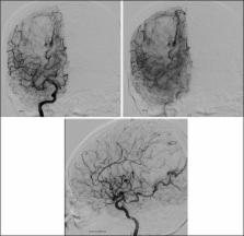
          We present a case of spontaneous obliteration of AVM in a 60-year-old gentleman who presented with intraparenchymal hemorrhage from a ruptured right parieto-occipital AVM. Angiography performed before gamma knife surgery 4 months after his initial presentation demonstrated complete absence of AVM.

          Conclusion:

          In our center's 20-year experience of treatment of cerebral AVMs (approximately 600 cases), this is the only case that has been aborted due to spontaneous obliteration leading us to infer that the incidence of spontaneous AVM obliteration is <1%. Spontaneous obliteration of AVM is a rare but well-established phenomenon that bears testimony to the dynamics of this vascular disorder.

          Related collections

          Most cited references27

          • Record: found
          • Abstract: found
          • Article: not found

          Embryological basis of some aspects of cerebral vascular fistulas and malformations.

          The literature on the formation of cerebral veins is reviewed to obtain a better understanding of some cerebrovascular anomalies. Clinical observations such as the entry of the superior ophthalmic vein into the cavernous sinus through the inferior rather than the superior orbital fissure, the relative infrequency of middle cerebral vein backflow in the presence of an extensive cavernous fistula, and the relative infrequency of hemorrhage in relation to the inferior petrosal fistula all relate to the persistence of an older venous pathway. The frequent occurrence of hemorrhage in association with the superior petrosal sinus fistula and the frequent failure of the superior petrosal sinus to connect to the cavernous sinus similarly have an embryological explanation. The frequent association of the vein of Galen aneurysm and an absent or deformed straight sinus probably relates to the time at which the paired internal cerebral veins fuse into one channel. It is speculated that the origins of cerebral venous malformations (CVMs) and arteriovenous malformations (AVMs) probably relate to sequential formation and absorption of surface veins, which occur in human embryonic development mainly in the 40- to 80-mm length interval, although persistent AVM growth is possible even after birth. The frequent absence or anomaly of the middle cerebral vein and its failure to communicate with the cavernous sinus in the presence of both CVMs and AVMs are linked to the late development of that vein and to its even later connection to the cavernous sinus.
            Bookmark
            • Record: found
            • Abstract: found
            • Article: not found

            Spontaneous angiographic obliteration of cerebral arteriovenous malformations.

            The factors associated with spontaneous angiographic obliteration of cerebral arteriovenous malformations (AVMs) are not well understood. We present a review of the literature and a report of our experience with six cases (four with no previous treatment intervention and two postoperative residual malformations) that were identified as having occurred during a 20-year period and describe the clinical and lesion features associated with this rare phenomenon. We present the first detailed histological study of a spontaneously thrombosed AVM specimen, including immunohistochemical analysis of angiogenesis factor expression. A combined experience in the management of approximately 700 AVMs during 20 years identified six cases of spontaneous angiographic obliteration of cerebral AVMs. A literature review revealed another 24 cases with angiographic documentation of the initial AVMs and follow-up data showing nonfilling of the lesions. Histological analysis of a recently excised lesion included immunostaining with monoclonal antibodies to the antigens of Factor VIII, Tie, vascular endothelial growth factor, and its receptors, Flt-1 and Flk. A single draining vein was a feature in each of our 6 cases and in 12 of 14 (86%) cases from the literature. Hemorrhage as the presenting symptom was identified in 5 of our 6 (83%) cases and in 17 of 24 (71%) of the literature cases. The size of the AVM was less than 6 cm in each of our 6 cases and in 22 of 24 (92%) of the literature cases. A histological examination of a thrombosed AVM surgical specimen revealed persistent patent vascular channels within the lesion. Immunohistochemical analysis with angiogenesis and endothelia-specific factors showed expression of these factors within the lumen of the thrombosed nidus vessels. We propose that the occlusion of a single draining vein may lead to total venous outflow obstruction and lesion thrombosis. Hemorrhagic presentation and small nidus may also predispose to this phenomenon. Immunohistochemical analysis of a thrombosed AVM revealed possible ongoing angiogenic changes within the AVM vessels 1 month after angiographically documented thrombosis. It is possible that neovascularization within a thrombosed AVM may lead to lesion recanalization; however, this phenomenon seems to be clinically exceedingly rare.
              Bookmark
              • Record: found
              • Abstract: found
              • Article: not found

              Cerebral arteriovenous malformations. Part 2: physiology.

              The scientific understanding of the nature of arteriovenous malformations (AVMs) in the brain is evolving. It is clear from current work that AVMs can undergo a variety of phenomena, including growth, remodeling, and/or regression-and the responsible processes are both molecular and physiological. A review of these complex processes is critical to directing future therapeutic approaches. The authors performed a comprehensive review of the literature to evaluate current information regarding the genetics, pathophysiology, and behavior of AVMs. A comprehensive literature review was conducted using PubMed to reveal the angioarchitecture and cerebral hemodynamics of AVMS as they relate to lesion development. Feeding artery pressures, brain AVM compartmentalization, venous drainage, flow phenomena, and vascular steal are discussed. The dynamic nature of brain AVMs is at least in part attributable to hemodynamic and flow-related phenomena. These forces acting on an evolving structure are critical to understanding the challenges in endovascular and surgical therapy. As knowledge in this field continues to progress, the natural history and predicted behavior of these AVMs will become more clearly elucidated.
                Bookmark

                Author and article information

                Contributors
                Journal
                Surg Neurol Int
                Surg Neurol Int
                SNI
                Surgical Neurology International
                Medknow Publications & Media Pvt Ltd (India )
                2229-5097
                2152-7806
                2016
                20 April 2016
                : 7
                : 45
                Affiliations
                [1]Department of Surgery, Division of Neurosurgery, National University Hospital, 5 Lower Kent Ridge Road, 119074 Singapore
                Author notes
                [* ]Corresponding author
                Article
                SNI-7-45
                10.4103/2152-7806.180766
                4854034
                27168948
                59f4c77a-ea70-4072-aa2b-caa278e7fbed
                Copyright: © 2016 Surgical Neurology International

                This is an open access article distributed under the terms of the Creative Commons Attribution-NonCommercial-ShareAlike 3.0 License, which allows others to remix, tweak, and build upon the work non-commercially, as long as the author is credited and the new creations are licensed under the identical terms.

                History
                : 16 September 2015
                : 07 February 2016
                Categories
                Case Report

                Surgery
                cerebral arteriovenous malformation,dynamic lesion,spontaneous obliteration
                Surgery
                cerebral arteriovenous malformation, dynamic lesion, spontaneous obliteration

                Comments

                Comment on this article