29
views
0
recommends
+1 Recommend
0 collections
    0
    shares
      • Record: found
      • Abstract: found
      • Article: found
      Is Open Access

      Genetically modified human placenta-derived mesenchymal stem cells with FGF-2 and PDGF-BB enhance neovascularization in a model of hindlimb ischemia

      research-article

      Read this article at

      Bookmark
          There is no author summary for this article yet. Authors can add summaries to their articles on ScienceOpen to make them more accessible to a non-specialist audience.

          Abstract

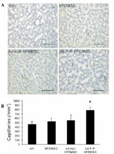
          Ischemic diseases represent a challenging worldwide health burden. The current study investigated the therapeutic potential of genetically modified human placenta-derived mesenchymal stem cells (hPDMSCs) with basic fibroblast growth factor (FGF2) and platelet-derived growth factor-BB (PDGF-BB) genes in hindlimb ischemia. Mesenchymal stem cells obtained from human term placenta were transfected ex vivo with adenoviral bicistronic vectors carrying the FGF2 and PDGF-BB genes (Ad-F-P). Unilateral hindlimb ischemia was surgically induced by excision of the right femoral artery in New Zealand White rabbits. Ad-F-P genetically modified hPDMSCs, Ad-null (control vector)-modified hPDMSCs, unmodified hPDMSCs or media were intramuscularly implanted into the ischemic limbs 7 days subsequent to the induction of ischemia. Four weeks after cell therapy, angiographic analysis revealed significantly increased collateral vessel formation in the Ad-F-P-hPDMSC group compared with the control group. Histological examination revealed markedly increased capillary and arteriole density in the Ad-F-P-hPDMSC group. The xenografted hPDMSCs survived in the ischemic tissue for at least 4 weeks subsequent to cell therapy. The current study demonstrated that the combination of hPDMSC therapy with FGF2 and PDGF-BB gene therapy effectively induced collateral vessel formation and angiogenesis, suggesting a novel strategy for therapeutic angiogenesis.

          Related collections

          Most cited references32

          • Record: found
          • Abstract: found
          • Article: not found

          Concise review: isolation and characterization of cells from human term placenta: outcome of the first international Workshop on Placenta Derived Stem Cells.

          Placental tissue draws great interest as a source of cells for regenerative medicine because of the phenotypic plasticity of many of the cell types isolated from this tissue. Furthermore, placenta, which is involved in maintaining fetal tolerance, contains cells that display immunomodulatory properties. These two features could prove useful for future cell therapy-based clinical applications. Placental tissue is readily available and easily procured without invasive procedures, and its use does not elicit ethical debate. Numerous reports describing stem cells from different parts of the placenta, using nearly as numerous isolation and characterization procedures, have been published. Considering the complexity of the placenta, an urgent need exists to define, as clearly as possible, the region of origin and methods of isolation of cells derived from this tissue. On March 23-24, 2007, the first international Workshop on Placenta Derived Stem Cells was held in Brescia, Italy. Most of the research published in this area focuses on mesenchymal stromal cells isolated from various parts of the placenta or epithelial cells isolated from amniotic membrane. The aim of this review is to summarize and provide the state of the art of research in this field, addressing aspects such as cell isolation protocols and characteristics of these cells, as well as providing preliminary indications of the possibilities for use of these cells in future clinical applications.
            Bookmark
            • Record: found
            • Abstract: found
            • Article: not found

            Local delivery of marrow-derived stromal cells augments collateral perfusion through paracrine mechanisms.

            Bone marrow cell therapy is reported to contribute to collateral formation through cell incorporation into new or remodeling vessels. However, the possible role of a paracrine contribution to this effect is less well characterized. Murine marrow-derived stromal cells (MSCs) were purified by magnetic bead separation of cultured bone marrow. The release of vascular endothelial growth factor (VEGF), basic fibroblast growth factor (bFGF), placental growth factor (PlGF), and monocyte chemoattractant protein-1 (MCP-1) was demonstrated by analysis of MSC conditioned media (MSC-CM). MSC-CM enhanced proliferation of endothelial cells and smooth muscle cells in a dose-dependent manner; anti-VEGF and anti-FGF antibodies only partly attenuated these effects. Balb/C mice (n=10) underwent distal femoral artery ligation, followed by adductor muscle injection of 1x10(6) MSCs 24 hours later. Compared with controls injected with media (n=10) or mature endothelial cells (n=8), distal limb perfusion improved, and mid-thigh conductance vessels increased in number and total cross-sectional area. MSC injection improved limb function and appearance, reduced the incidence of auto-amputation, and attenuated muscle atrophy and fibrosis. After injection, labeled MSCs were seen dispersed between muscle fibers but were not seen incorporated into mature collaterals. Injection of MSCs increased adductor muscle levels of bFGF and VEGF protein compared with controls. Finally, colocalization of VEGF and transplanted MSCs within adductor tissue was demonstrated. MSCs secrete a wide array of arteriogenic cytokines. MSCs can contribute to collateral remodeling through paracrine mechanisms.
              Bookmark
              • Record: found
              • Abstract: found
              • Article: not found

              Human mesenchymal stem cells engraft and demonstrate site-specific differentiation after in utero transplantation in sheep.

              Mesenchymal stem cells are multipotent cells that can be isolated from adult bone marrow and can be induced in vitro and in vivo to differentiate into a variety of mesenchymal tissues, including bone, cartilage, tendon, fat, bone marrow stroma, and muscle. Despite their potential clinical utility for cellular and gene therapy, the fate of mesenchymal stem cells after systemic administration is mostly unknown. To address this, we transplanted a well-characterized human mesenchymal stem cell population into fetal sheep early in gestation, before and after the expected development of immunologic competence. In this xenogeneic system, human mesenchymal stem cells engrafted and persisted in multiple tissues for as long as 13 months after transplantation. Transplanted human cells underwent site-specific differentiation into chondrocytes, adipocytes, myocytes and cardiomyocytes, bone marrow stromal cells and thymic stroma. Unexpectedly, there was long-term engraftment even when cells were transplanted after the expected development of immunocompetence. Thus, mesenchymal stem cells maintain their multipotential capacity after transplantation, and seem to have unique immunologic characteristics that allow persistence in a xenogeneic environment. Our data support the possibility of the transplantability of mesenchymal stem cells and their potential utility in tissue engineering, and cellular and gene therapy applications.
                Bookmark

                Author and article information

                Journal
                Mol Med Rep
                Mol Med Rep
                Molecular Medicine Reports
                D.A. Spandidos
                1791-2997
                1791-3004
                October 2015
                20 July 2015
                20 July 2015
                : 12
                : 4
                : 5093-5099
                Affiliations
                [1 ]State Key Laboratory of Biotherapy and Cancer Center, West China Hospital, Sichuan University, and Collaborative Innovation Center for Biotherapy, Chengdu, Sichuan 610041, P.R. China
                [2 ]Department of Thoracic Oncology, Cancer Center, West China Hospital, Sichuan University, Chengdu, Sichuan 610041, P.R. China
                Author notes
                Correspondence to: Dr Li Yang or Dr Yuquan Wei, State Key Laboratory of Biotherapy and Cancer Center, West China Hospital, Sichuan University, and Collaborative Innovation Center for Biotherapy, 17 People's South Road, Chengdu, Sichuan 610041, P.R. China, E-mail: yl.tracy73@ 123456gmail.com , E-mail: yqwei@ 123456vip.sina.com
                [*]

                Contributed equally

                Article
                mmr-12-04-5093
                10.3892/mmr.2015.4089
                4581748
                26239842
                59f686e4-2f69-4639-9eaa-6e811a5ffa1e
                Copyright: © Yin.

                This is an open access article distributed under the terms of a Creative Commons Attribution License

                History
                : 01 December 2013
                : 26 June 2015
                Categories
                Articles

                ischemia,angiogenesis,arteriogenesis,gene therapy
                ischemia, angiogenesis, arteriogenesis, gene therapy

                Comments

                Comment on this article