0
views
0
recommends
+1 Recommend
0 collections
    0
    shares
      • Record: found
      • Abstract: found
      • Article: found
      Is Open Access

      Clinical features and treatment response to differentiate idiopathic peritonitis from non-strangulating intestinal infarction of the pelvic flexure associated with Strongylus vulgaris infection in the horse

      research-article

      Read this article at

      Bookmark
          There is no author summary for this article yet. Authors can add summaries to their articles on ScienceOpen to make them more accessible to a non-specialist audience.

          Abstract

          Background

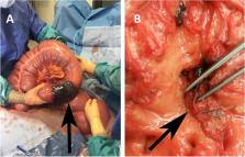
          Peritonitis in horses secondary to non-strangulating infarction (NSII) has a guarded prognosis, even after intestinal resection. In contrast, horses with idiopathic peritonitis respond well to medical treatment. Affected horses in both cases often show signs of both colic and systemic inflammation, but early diagnosis is crucial for optimal treatment and an accurate prognosis. One cause of NSII is thrombus formation secondary to Strongylus vulgaris larval migration. There has been a documented increase in S. vulgaris prevalence in Sweden since the implementation of selective anthelmintic treatment in 2007, which subsequently could result in a rise in NSII cases. In a retrospective clinical study, medical records from cases diagnosed with NSII of the pelvic flexure or idiopathic peritonitis from three equine referral hospitals in Sweden during 2017–2020 were reviewed. Information including demographic data, relevant medical history, and clinical- and laboratory parameters were obtained from patient records. To facilitate the differentiation between cases of idiopathic peritonitis and cases with confirmed NSII of the pelvic flexure, the aim of the study was to compare clinical and laboratory parameters, clinical progression and initial response to antimicrobial treatment. A secondary aim was to compare survival-rates.

          Results

          Horses with NSII ( n = 20) were significantly more likely to present during the winter months with a poorer response to medical treatment within 48 h. Cases of idiopathic peritonitis ( n = 107) had a 100% survival rate with medical treatment, although one case required surgical correction of a colon displacement. In comparison, all confirmed NSII cases were non-responsive to antimicrobial treatment, with a survival rate to discharge of 50% after colon resection. Specific rectal findings and peripheral blood neutropenia were strongly associated with NSII.

          Conclusions

          In Sweden, idiopathic peritonitis cases still predominate over S. vulgaris associated NSII cases and have an excellent survival rate with antimicrobial treatment. However, horses presenting with septic peritonitis during the winter months with a palpable rectal mass and displaying fever and colic signs beyond 48 h of medical treatment are likely to suffer from NSII of the pelvic flexure and should be considered for abdominal surgery.

          Supplementary Information

          The online version contains supplementary material available at 10.1186/s12917-022-03248-x.

          Related collections

          Most cited references35

          • Record: found
          • Abstract: found
          • Article: not found

          World Association for the Advancement of Veterinary Parasitology (W.A.A.V.P.) methods for the detection of anthelmintic resistance in nematodes of veterinary importance.

          Methods have been described to assist in the detection of anthelmintic resistance in strongylid nematodes of ruminants, horses and pigs. Two tests are recommended, an in vivo test, the faecal egg count reduction test for use in infected animals, and an in vitro test, the egg hatch test for detection of benzimidazole resistance in nematodes that hatch shortly after embryonation. Anaerobic storage for submission of faecal samples from the field for use in the in vitro test is of value and the procedure is described. The tests should enable comparable data to be obtained in surveys in all parts of the world.
            Bookmark
            • Record: found
            • Abstract: not found
            • Book: not found

            Modern Applied Statistics with S

              Bookmark
              • Record: found
              • Abstract: found
              • Article: not found

              Actinobacillus species and their role in animal disease.

              Actinobacillus species are Gram-negative bacteria responsible for several quite distinct disease conditions of animals. The natural habitat of the organisms is primarily the upper respiratory tract and oral cavity. A. lignieresii is the cause of actinomycosis (wooden tongue) in cattle: a sporadic, insidiously-developing granulomatous infection. In sharp contrast is A. pleuropneumoniae which is responsible for a rapidly spreading often fatal pneumonia, common among intensively reared pigs. Detailed investigation of this organism has provided a much clearer picture of the bacterial factors involved in causing disease. A. equuli similarly causes a potent septicaemia in the neonatal foal; growing apparently unrestricted once infection occurs. Other members of the genus induce characteristic pathogenesis in their preferred host, with one, A. actinomycetemcomitans, being a cause of human periodontal disease. This article reviews recent understanding of the taxonomy and bacteriology of the organisms, and the aetiology, pathogenicity, diagnosis and control of animal disease caused by Actinobacillus species. Copyright 2000 Harcourt Publishers Ltd.
                Bookmark

                Author and article information

                Contributors
                ylva.hedberg.alm@uds.slu.se
                Journal
                BMC Vet Res
                BMC Vet Res
                BMC Veterinary Research
                BioMed Central (London )
                1746-6148
                23 April 2022
                23 April 2022
                2022
                : 18
                : 149
                Affiliations
                [1 ]GRID grid.6341.0, ISNI 0000 0000 8578 2742, Department of Biomedical Science and Veterinary Public Health, Parasitology Unit, , Swedish University of Agricultural Sciences, ; Uppsala, Sweden
                [2 ]GRID grid.6341.0, ISNI 0000 0000 8578 2742, Equine Clinic, University Animal Hospital, Swedish University of Agricultural Sciences, ; Uppsala, Sweden
                [3 ]GRID grid.6341.0, ISNI 0000 0000 8578 2742, Department of Clinical Sciences, Veterinary Epidemiology Unit, , Swedish University of Agricultural Sciences, ; Uppsala, Sweden
                [4 ]GRID grid.6341.0, ISNI 0000 0000 8578 2742, Department of Biomedical Science and Veterinary Public Health, Pathology Unit, , Swedish University of Agricultural Sciences, ; Uppsala, Sweden
                [5 ]Specialist Equine Hospital, Evidensia Animal Hospital, Helsingborg, Sweden
                [6 ]GRID grid.6341.0, ISNI 0000 0000 8578 2742, Department of Clinical Sciences, Equine Medicine Unit, , Swedish University of Agricultural Sciences, ; Uppsala, Sweden
                Article
                3248
                10.1186/s12917-022-03248-x
                9034621
                35461295
                5a834ddf-4b4a-4897-bac5-1bba84775579
                © The Author(s) 2022

                Open AccessThis article is licensed under a Creative Commons Attribution 4.0 International License, which permits use, sharing, adaptation, distribution and reproduction in any medium or format, as long as you give appropriate credit to the original author(s) and the source, provide a link to the Creative Commons licence, and indicate if changes were made. The images or other third party material in this article are included in the article's Creative Commons licence, unless indicated otherwise in a credit line to the material. If material is not included in the article's Creative Commons licence and your intended use is not permitted by statutory regulation or exceeds the permitted use, you will need to obtain permission directly from the copyright holder. To view a copy of this licence, visit http://creativecommons.org/licenses/by/4.0/. The Creative Commons Public Domain Dedication waiver ( http://creativecommons.org/publicdomain/zero/1.0/) applies to the data made available in this article, unless otherwise stated in a credit line to the data.

                History
                : 12 October 2021
                : 12 April 2022
                Funding
                Funded by: The Swedish-Norwegian Foundation for Veterinary Research
                Funded by: Swedish University of Agricultural Sciences
                Categories
                Research
                Custom metadata
                © The Author(s) 2022

                Veterinary medicine
                equine,intestinal parasite,colic,rectal examination,prognosis
                Veterinary medicine
                equine, intestinal parasite, colic, rectal examination, prognosis

                Comments

                Comment on this article