4
views
0
recommends
+1 Recommend
0 collections
    0
    shares
      • Record: found
      • Abstract: found
      • Article: found
      Is Open Access

      Mutant ASXL1 cooperates with BAP1 to promote myeloid leukaemogenesis

      research-article

      Read this article at

      Bookmark
          There is no author summary for this article yet. Authors can add summaries to their articles on ScienceOpen to make them more accessible to a non-specialist audience.

          Abstract

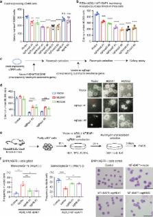
          ASXL1 mutations occur frequently in myeloid neoplasms and are associated with poor prognosis. However, the mechanisms by which mutant ASXL1 induces leukaemogenesis remain unclear. In this study, we report mutually reinforcing effects between a C-terminally truncated form of mutant ASXL1 (ASXL1-MT) and BAP1 in promoting myeloid leukaemogenesis. BAP1 expression results in increased monoubiquitination of ASXL1-MT, which in turn increases the catalytic function of BAP1. This hyperactive ASXL1-MT/BAP1 complex promotes aberrant myeloid differentiation of haematopoietic progenitor cells and accelerates RUNX1-ETO-driven leukaemogenesis. Mechanistically, this complex induces upregulation of posterior HOXA genes and IRF8 through removal of H2AK119 ubiquitination. Importantly, BAP1 depletion inhibits posterior HOXA gene expression and leukaemogenicity of ASXL1-MT-expressing myeloid leukemia cells. Furthermore, BAP1 is also required for the growth of MLL-fusion leukemia cells with posterior HOXA gene dysregulation. These data indicate that BAP1, which has long been considered a tumor suppressor, in fact plays tumor-promoting roles in myeloid neoplasms.

          Abstract

          ASXL1 gene is often mutated in myeloid malignancies. Here, the authors show that mutant ASXL1 and BAP1 are in a positive feedback loop such that BAP1 induces monoubiquitination of mutant ASXL1, which in turn enhances BAP1 activity to potentiate myeloid transformation via HOXA clusters and IRF8.

          Related collections

          Most cited references43

          • Record: found
          • Abstract: found
          • Article: not found

          Clinical effect of point mutations in myelodysplastic syndromes.

          Myelodysplastic syndromes are clinically heterogeneous disorders characterized by clonal hematopoiesis, impaired differentiation, peripheral-blood cytopenias, and a risk of progression to acute myeloid leukemia. Somatic mutations may influence the clinical phenotype but are not included in current prognostic scoring systems. We used a combination of genomic approaches, including next-generation sequencing and mass spectrometry-based genotyping, to identify mutations in samples of bone marrow aspirate from 439 patients with myelodysplastic syndromes. We then examined whether the mutation status for each gene was associated with clinical variables, including specific cytopenias, the proportion of blasts, and overall survival. We identified somatic mutations in 18 genes, including two, ETV6 and GNAS, that have not been reported to be mutated in patients with myelodysplastic syndromes. A total of 51% of all patients had at least one point mutation, including 52% of the patients with normal cytogenetics. Mutations in RUNX1, TP53, and NRAS were most strongly associated with severe thrombocytopenia (P<0.001 for all comparisons) and an increased proportion of bone marrow blasts (P<0.006 for all comparisons). In a multivariable Cox regression model, the presence of mutations in five genes retained independent prognostic significance: TP53 (hazard ratio for death from any cause, 2.48; 95% confidence interval [CI], 1.60 to 3.84), EZH2 (hazard ratio, 2.13; 95% CI, 1.36 to 3.33), ETV6 (hazard ratio, 2.04; 95% CI, 1.08 to 3.86), RUNX1 (hazard ratio, 1.47; 95% CI, 1.01 to 2.15), and ASXL1 (hazard ratio, 1.38; 95% CI, 1.00 to 1.89). Somatic point mutations are common in myelodysplastic syndromes and are associated with specific clinical features. Mutations in TP53, EZH2, ETV6, RUNX1, and ASXL1 are predictors of poor overall survival in patients with myelodysplastic syndromes, independently of established risk factors. (Funded by the National Institutes of Health and others.).
            Bookmark
            • Record: found
            • Abstract: found
            • Article: not found

            Histone H2A deubiquitinase activity of the Polycomb repressive complex PR-DUB.

            Polycomb group (PcG) proteins are transcriptional repressors that control processes ranging from the maintenance of cell fate decisions and stem cell pluripotency in animals to the control of flowering time in plants. In Drosophila, genetic studies identified more than 15 different PcG proteins that are required to repress homeotic (HOX) and other developmental regulator genes in cells where they must stay inactive. Biochemical analyses established that these PcG proteins exist in distinct multiprotein complexes that bind to and modify chromatin of target genes. Among those, Polycomb repressive complex 1 (PRC1) and the related dRing-associated factors (dRAF) complex contain an E3 ligase activity for monoubiquitination of histone H2A (refs 1-4). Here we show that the uncharacterized Drosophila PcG gene calypso encodes the ubiquitin carboxy-terminal hydrolase BAP1. Biochemically purified Calypso exists in a complex with the PcG protein ASX, and this complex, named Polycomb repressive deubiquitinase (PR-DUB), is bound at PcG target genes in Drosophila. Reconstituted recombinant Drosophila and human PR-DUB complexes remove monoubiquitin from H2A but not from H2B in nucleosomes. Drosophila mutants lacking PR-DUB show a strong increase in the levels of monoubiquitinated H2A. A mutation that disrupts the catalytic activity of Calypso, or absence of the ASX subunit abolishes H2A deubiquitination in vitro and HOX gene repression in vivo. Polycomb gene silencing may thus entail a dynamic balance between H2A ubiquitination by PRC1 and dRAF, and H2A deubiquitination by PR-DUB.
              Bookmark
              • Record: found
              • Abstract: found
              • Article: not found

              ASXL1 mutations promote myeloid transformation through loss of PRC2-mediated gene repression.

              Recurrent somatic ASXL1 mutations occur in patients with myelodysplastic syndrome, myeloproliferative neoplasms, and acute myeloid leukemia, and are associated with adverse outcome. Despite the genetic and clinical data implicating ASXL1 mutations in myeloid malignancies, the mechanisms of transformation by ASXL1 mutations are not understood. Here, we identify that ASXL1 mutations result in loss of polycomb repressive complex 2 (PRC2)-mediated histone H3 lysine 27 (H3K27) tri-methylation. Through integration of microarray data with genome-wide histone modification ChIP-Seq data, we identify targets of ASXL1 repression, including the posterior HOXA cluster that is known to contribute to myeloid transformation. We demonstrate that ASXL1 associates with the PRC2, and that loss of ASXL1 in vivo collaborates with NRASG12D to promote myeloid leukemogenesis. Copyright © 2012 Elsevier Inc. All rights reserved.
                Bookmark

                Author and article information

                Contributors
                kitamura@ims.u-tokyo.ac.jp
                Journal
                Nat Commun
                Nat Commun
                Nature Communications
                Nature Publishing Group UK (London )
                2041-1723
                16 July 2018
                16 July 2018
                2018
                : 9
                : 2733
                Affiliations
                [1 ]ISNI 0000 0001 2151 536X, GRID grid.26999.3d, Division of Cellular Therapy, Advanced Clinical Research Center, and Division of Stem Cell Signaling, Center for Stem Cell Biology and Regenerative Medicine, Institute of Medical Science, , The University of Tokyo, ; Tokyo, 1088639 Japan
                [2 ]ISNI 0000 0001 2171 9952, GRID grid.51462.34, Human Oncology and Pathogenesis Program, , Memorial Sloan Kettering Cancer Center, ; New York, NY 10065 USA
                [3 ]ISNI 000000041936877X, GRID grid.5386.8, Department of Hematology/Oncology, , Weill Cornell Medical College, ; New York, NY 10021 USA
                [4 ]National Cancer Center Tsuruoka Metabolomics Laboratory, Yamagata, 9970052 Japan
                [5 ]ISNI 0000 0001 2151 536X, GRID grid.26999.3d, Division of Stem Cell Therapy, The Institute of Medical Science, , The University of Tokyo, ; Tokyo, 1088639 Japan
                [6 ]ISNI 0000 0001 2151 536X, GRID grid.26999.3d, Medical Proteomics Laboratory, The Institute of Medical Science, , The University of Tokyo, ; Tokyo, 1088639 Japan
                [7 ]ISNI 0000 0001 2151 536X, GRID grid.26999.3d, Department of Cellular Signaling, Graduate School of Medicine, , The University of Tokyo, ; Tokyo, 1130033 Japan
                [8 ]ISNI 0000 0001 2151 536X, GRID grid.26999.3d, Department of Medical Genomics, Graduate School of Medicine, , The University of Tokyo, ; Tokyo, 1130033 Japan
                [9 ]ISNI 0000 0001 2168 5385, GRID grid.272242.3, National Cancer Center Research Institute, ; Tokyo, 1040045 Japan
                Author information
                http://orcid.org/0000-0003-4146-3629
                Article
                5085
                10.1038/s41467-018-05085-9
                6048047
                30013160
                5a9b7c6a-8fd3-47af-a7f2-f4ab96943b90
                © The Author(s) 2018

                Open Access This article is licensed under a Creative Commons Attribution 4.0 International License, which permits use, sharing, adaptation, distribution and reproduction in any medium or format, as long as you give appropriate credit to the original author(s) and the source, provide a link to the Creative Commons license, and indicate if changes were made. The images or other third party material in this article are included in the article’s Creative Commons license, unless indicated otherwise in a credit line to the material. If material is not included in the article’s Creative Commons license and your intended use is not permitted by statutory regulation or exceeds the permitted use, you will need to obtain permission directly from the copyright holder. To view a copy of this license, visit http://creativecommons.org/licenses/by/4.0/.

                History
                : 2 November 2017
                : 13 June 2018
                Categories
                Article
                Custom metadata
                © The Author(s) 2018

                Uncategorized
                Uncategorized

                Comments

                Comment on this article