4
views
0
recommends
+1 Recommend
0 collections
    0
    shares
      • Record: found
      • Abstract: found
      • Article: found
      Is Open Access

      Systemically administered neurotensin receptor agonist produces antinociception through activation of spinally projecting serotonergic neurons in the rostral ventromedial medulla

      research-article

      Read this article at

      Bookmark
          There is no author summary for this article yet. Authors can add summaries to their articles on ScienceOpen to make them more accessible to a non-specialist audience.

          Abstract

          Background

          Supraspinal delivery of neurotensin (NTS), which may contribute to the effect of a systemically administered agonist, has been reported to be either pronociceptive or antinociceptive. Here, we evaluated the effects of systemically administered NTSR1 agonist in a rat model of neuropathic pain and elucidated the underlying supraspinal mechanism.

          Methods

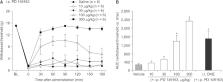
          Neuropathic pain was induced by L5 and L6 spinal nerve ligation in male Sprague–Dawley rats. The effects of intraperitoneally administered NTSR1 agonist PD 149163 was assessed using von Frey filaments. To examine the role of 5-HT neurotransmission, a serotonin (5-HT) receptor antagonist dihydroergocristine was pretreated intrathecally, and spinal microdialysis studies were performed to measure the change in extracellular level of 5-HT in response to PD 149163 administration. To investigate the supraspinal mechanism, NTSR1 antagonist 48692 was microinjected into the rostral ventromedial medulla (RVM) prior to systemic PD 149163. Additionally, the effect of intrathecal DHE on intra-RVM PD 149163 was assessed.

          Results

          Intraperitoneally administered PD 149163 exhibited a dose-dependent attenuation of mechanical allodynia. This effect was partially reversed by intrathecal pretreatment with dihydroergocristine and was accompanied by an increased extracellular level of 5-HT in the spinal cord. The PD 149163-produced antinociception was also blocked by intra-RVM SB 48692. Direct injection of PD 149163 into the RVM mimicked the maximum effect of the same drug delivered intraperitoneally, which was reversed by intrathecal dihydroergocristine.

          Conclusions

          These observations indicate that systemically administered NTSR1 agonist produces antinociception through the NTSR1 in the RVM, activating descending serotonergic projection to release 5-HT into the spinal dorsal horn.

          Related collections

          Most cited references26

          • Record: found
          • Abstract: found
          • Article: not found

          Quantitative assessment of tactile allodynia in the rat paw

          We applied and validated a quantitative allodynia assessment technique, using a recently developed rat surgical neuropathy model wherein nocifensive behaviors are evoked by light touch to the paw. Employing von Frey hairs from 0.41 to 15.1 g, we first characterized the percent response at each stimulus intensity. A smooth log-linear relationship was observed, with a median 50% threshold at 1.97 g (95% confidence limits, 1.12-3.57 g). Subsequently, we applied a paradigm using stimulus oscillation around the response threshold, which allowed more rapid, efficient measurements. Median 50% threshold by this up-down method was 2.4 g (1.81-2.76). Correlation coefficient between the two methods was 0.91. In neuropathic rats, good intra- and inter-observer reproducibility was found for the up-down paradigm; some variability was seen in normal rats, attributable to extensive testing. Thresholds in a sizable group of neuropathic rats showed insignificant variability over 20 days. After 50 days, 61% still met strict neuropathy criteria, using survival analysis. Threshold measurement using the up-down paradigm, in combination with the neuropathic pain model, represents a powerful tool for analyzing the effects of manipulations of the neuropathic pain state.
            Bookmark
            • Record: found
            • Abstract: found
            • Article: not found

            Descending control of pain.

            Upon receipt in the dorsal horn (DH) of the spinal cord, nociceptive (pain-signalling) information from the viscera, skin and other organs is subject to extensive processing by a diversity of mechanisms, certain of which enhance, and certain of which inhibit, its transfer to higher centres. In this regard, a network of descending pathways projecting from cerebral structures to the DH plays a complex and crucial role. Specific centrifugal pathways either suppress (descending inhibition) or potentiate (descending facilitation) passage of nociceptive messages to the brain. Engagement of descending inhibition by the opioid analgesic, morphine, fulfils an important role in its pain-relieving properties, while induction of analgesia by the adrenergic agonist, clonidine, reflects actions at alpha(2)-adrenoceptors (alpha(2)-ARs) in the DH normally recruited by descending pathways. However, opioids and adrenergic agents exploit but a tiny fraction of the vast panoply of mechanisms now known to be involved in the induction and/or expression of descending controls. For example, no drug interfering with descending facilitation is currently available for clinical use. The present review focuses on: (1) the organisation of descending pathways and their pathophysiological significance; (2) the role of individual transmitters and specific receptor types in the modulation and expression of mechanisms of descending inhibition and facilitation and (3) the advantages and limitations of established and innovative analgesic strategies which act by manipulation of descending controls. Knowledge of descending pathways has increased exponentially in recent years, so this is an opportune moment to survey their operation and therapeutic relevance to the improved management of pain.
              Bookmark
              • Record: found
              • Abstract: found
              • Article: not found

              An experimental model for peripheral neuropathy produced by segmental spinal nerve ligation in the rat

              We attempted to develop an experimental animal model for peripheral neuropathic pain. Under sodium pentobarbital anesthesia, both the L5 and L6 spinal nerves (group 1) or the L5 spinal nerve alone (group 2) of one side of the rat were tightly ligated. For comparison, a parallel study was conducted with another group of rats (group 3) which received a partial tight sciatic nerve ligation, a paradigm developed previously as a neuropathy model. Withdrawal latencies to application of radiant heat to the foot were tested for the next 16 weeks in all 3 groups. Sensitivity of the hind paw to mechanical stimulation was tested with von Frey filaments. The general behavior of each rat was noted during the entire test period. Results suggested that the surgical procedure in all 3 groups produced a long-lasting hyperalgesia to noxious heat (at least 5 weeks) and mechanical allodynia (at least 10 weeks) of the affected foot. In addition, there were behavioral signs of the presence of spontaneous pain in the affected foot. Therefore, we believe we have developed an experimental animal model for peripheral neuropathy using tight ligations of spinal nerves. The model manifests the symptoms of human patients with causalgia and is compatible with a previously developed neuropathy model. The present model has two unique features. First, the surgical procedure is stereotyped. Second, the levels of injured and intact spinal segments are completely separated, allowing independent experimental manipulations of the injured and intact spinal segments in future experiments to answer questions regarding mechanisms underlying causalgia.
                Bookmark

                Author and article information

                Journal
                Korean J Pain
                Korean J Pain
                The Korean Journal of Pain
                The Korean Pain Society
                2005-9159
                2093-0569
                1 January 2021
                1 January 2021
                : 34
                : 1
                : 58-65
                Affiliations
                [1 ]Department of Anesthesiology and Pain Medicine, Chonnam National University Hospital, Chonnam National University Medical School, Gwangju, Korea
                [2 ]Department of Anesthesiology and Pain Medicine, Chonnam National University Hwasun Hospital, Hwasun, Korea
                [3 ]Center for Creative Biomedical Scientists, Chonnam National University Medical School, Gwangju, Korea
                Author notes
                Correspondence Woong Mo Kim, Department of Anesthesiology and Pain Medicine, Chonnam National University Medical School, 160 Baekseo-ro, Dong-gu, Gwangju 61469, Korea, Tel: +82-62-220-6895, Fax: +82-62-232-6294, E-mail: kimwm@ 123456jnu.ac.kr , Hyung Gon Lee, Department of Anesthesiology and Pain Medicine, Chonnam National University Medical School, 160 Baekseo-ro, Dong-gu, Gwangju 61469, Korea, Tel: +82-62-220-6895, Fax: +82-62-232-6294, E-mail: leehg@ 123456jnu.ac.kr

                Handling Editor: Younghoon Jeon

                [*]

                These authors contributed equally to this work.

                Author information
                https://orcid.org/0000-0002-3073-0165
                https://orcid.org/0000-0002-5299-6861
                https://orcid.org/0000-0002-3523-7468
                https://orcid.org/0000-0003-4898-4355
                https://orcid.org/0000-0003-0579-6522
                https://orcid.org/0000-0002-3307-8563
                https://orcid.org/0000-0002-3571-2599
                https://orcid.org/0000-0002-9962-0255
                Article
                kjp-34-1-58
                10.3344/kjp.2021.34.1.58
                7783846
                33380568
                5b7da877-fef7-4490-b2e4-deb5b2b807e2
                © The Korean Pain Society, 2021

                This is an open-access article distributed under the terms of the Creative Commons Attribution Non-Commercial License ( http://creativecommons.org/licenses/by-nc/4.0), which permits unrestricted non-commercial use, distribution, and reproduction in any medium, provided the original work is properly cited.

                History
                : 28 October 2020
                : 12 December 2020
                : 16 December 2020
                Categories
                Experimental Research Articles

                Anesthesiology & Pain management
                analgesia,central nervous system,microdialysis,neuralgia,neurotensin,rats,receptors, neurotensin,serotonin,spinal cord

                Comments

                Comment on this article