1
views
0
recommends
+1 Recommend
0 collections
    0
    shares
      • Record: found
      • Abstract: found
      • Article: found
      Is Open Access

      Electrocardiographic changes in Emphysema

      review-article

      Read this article at

      Bookmark
          There is no author summary for this article yet. Authors can add summaries to their articles on ScienceOpen to make them more accessible to a non-specialist audience.

          Abstract

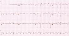
          Chronic obstructive lung disease (COPD), predominantly emphysema, causes several thoracic anatomical and hemodynamic changes which may cause changes in various electrocardiographic parameters. A 12-lead electrocardiogram (ECG), which is often a part of routine evaluation in most clinical settings, may serve as a useful screening modality for diagnosis of COPD or emphysema. Our current article aims to provide a comprehensive review of the electrocardiographic changes encountered in COPD/emphysema utilizing published PubMed and Medline literature database. Several important ECG changes are present in COPD/emphysema and may serve as a good diagnostic tool. Verticalization of P-vector, changes in QRS duration, pattern recognition of precordial R-wave progression and axial shifts can be considered some of the most valuable markers among other changes. In conclusion, 12-lead surface electrocardiogram can serve as a valuable tool for the diagnosis of COPD and/or emphysema. An appropriate knowledge of these ECG changes can not only help in the diagnosis but can also immensely help in an appropriate clinical management of these patients.

          Related collections

          Most cited references36

          • Record: found
          • Abstract: found
          • Article: not found

          Interatrial blocks. A separate entity from left atrial enlargement: a consensus report.

          Impaired interatrial conduction or interatrial block is well documented but is not described as an individual electrocardiographic (ECG) pattern in most of ECG books, although the term atrial abnormalities to encompass both concepts, left atrial enlargement (LAE) and interatrial block, has been coined. In fact, LAE and interatrial block are often associated, similarly to what happens with ventricular enlargement and ventricular block. The interatrial blocks, that is, the presence of delay of conduction between the right and left atria, are the most frequent atrial blocks. These may be of first degree (P-wave duration >120 milliseconds), third degree (longer P wave with biphasic [±] morphology in inferior leads), and second degree when these patterns appear transiently in the same ECG recording (atrial aberrancy). There are evidences that these electrocardiographic P-wave patterns are due to a block because they may (a) appear transiently, (b) be without associated atrial enlargement, and (c) may be reproduced experimentally. The presence of interatrial blocks may be seen in the absence of atrial enlargement but often are present in case of LAE. The most important clinical implications of interatrial block are the following: (a) the first degree interatrial blocks are very common, and their relation with atrial fibrillation and an increased risk for global and cardiovascular mortality has been demonstrated; (b) the third degree interatrial blocks are less frequent but are strong markers of LAE and paroxysmal supraventricular tachyarrhythmias. Their presence has been considered a true arrhythmological syndrome. Copyright © 2012 Elsevier Inc. All rights reserved.
            Bookmark
            • Record: found
            • Abstract: found
            • Article: not found

            Deaths: final data for 2008.

            This report presents final 2008 data on U.S. deaths, death rates, life expectancy, infant mortality, and trends by selected characteristics such as age, sex, Hispanic origin, race, state of residence, and cause of death. Information reported on death certificates, which is completed by funeral directors, attending physicians, medical examiners, and coroners, is presented in descriptive tabulations. The original records are filed in state registration offices. Statistical information is compiled in a national database through the Vital Statistics Cooperative Program of the Centers for Disease Control and Prevention's National Center for Health Statistics. Causes of death are processed in accordance with the International Classification of Diseases, Tenth Revision. In 2008, a total of 2,471,984 deaths were reported in the United States. The age-adjusted death rate was 758.3 deaths per 100,000 standard population, a decrease of 0.2 percent from the 2007 rate and a record low figure. Life expectancy at birth rose 0.2 years, from 77.9 years in 2007 to a record high 78.1 years in 2008. The age-specific death rate increased for age group 85 years and over. Age-specific death rates decreased for age groups: less than 1 year, 5-14, 15-24, 25-34, 35-44, and 65-74 years. The age-specific death rates remained unchanged for age groups: 1-4, 45-54, 55-64, and 75-84 years. The 15 leading causes of death in 2008 remained the same as in 2007, but Chronic lower respiratory diseases and suicide increased in the ranking while stroke and septicemia decreased in the ranking. Stroke is the fourth leading cause of death in 2008 after more than five decades at number three in the ranking. Chronic lower respiratory diseases is the third leading cause of death for 2008. The infant mortality rate decreased 2.1 percent to a historically low value of 6.61 deaths per 1000 live births in 2008. The decline of the age-adjusted death rate to a record low value for the United States and the increase in life expectancy to a record high value of 78.1 years are consistent with long-term trends in mortality.
              Bookmark
              • Record: found
              • Abstract: found
              • Article: not found

              Bayés' syndrome: the association between interatrial block and supraventricular arrhythmias.

              The past few years have given rise to extensive research on atrial conduction disorders and their clinical relevance. Most notably, an association between interatrial block and supraventricular arrhythmias has been discovered. This disorder, recently termed 'Bayés' syndrome', has important clinical implications. In this article, the authors review normal atrial conduction and associated disorders. A particular focus is placed on Bayés' syndrome and the relationship between interatrial block and supraventricular arrhythmias in different clinical scenarios. The report also outlines the current progress in the study of this syndrome and highlights areas requiring further investigation.
                Bookmark

                Author and article information

                Contributors
                Journal
                World J Cardiol
                WJC
                World Journal of Cardiology
                Baishideng Publishing Group Inc
                1949-8462
                26 October 2021
                26 October 2021
                : 13
                : 10
                : 533-545
                Affiliations
                Department of Interventional Cardiology, Northeast Ohio Medical University, Canton, OH 44272, United States
                BA-MD, Brooklyn College, Brooklyn, NY 11210, United States
                Department of Family Medicine, Memorial Hospital of Carbondale, Carbondale, IL 62901, United States
                Psychiatry, Loretto Hospital, Chicago, IL 60644, United States
                Department of Interventional Cardiology, St. Francis Medical Center, Monroe, LA 71201, United States
                Department of Cardiology, Westchester Medical Center Network Advanced Physician Services, Poughkeepsie, NY 12601, United States
                Department of Cardiology, Westchester Medical Center Network Advanced Physician Services, Poughkeepsie, NY 12601, United States. lovids@ 123456hotmail.com
                Author notes

                Author contributions: Gupta P performed the research and a major contribution to the manuscript draft; Jain H contributed to the manuscript draft, schematic illustrations copyright approvals and minor revisions; Gill M and Bharaj G contributed to draft revisions; Khalid N and Waseem C contributed to the analysis and revisions; Chhabra L conceptualized the project, supervised the research, contributed substantially to the manuscript draft, and performed major revisions.

                Corresponding author: Lovely Chhabra, FACC, MD, Doctor, Department of Cardiology, Westchester Medical Center Network Advanced Physician Services, 241 North Road, Poughkeepsie, NY 12601, United States. lovids@ 123456hotmail.com

                Article
                jWJC.v13.i10.pg533
                10.4330/wjc.v13.i10.533
                8554360
                34754398
                5e59efea-270a-4eb2-95d3-5fbeb8767881
                ©The Author(s) 2021. Published by Baishideng Publishing Group Inc. All rights reserved.

                This article is an open-access article that was selected by an in-house editor and fully peer-reviewed by external reviewers. It is distributed in accordance with the Creative Commons Attribution NonCommercial (CC BY-NC 4.0) license, which permits others to distribute, remix, adapt, build upon this work non-commercially, and license their derivative works on different terms, provided the original work is properly cited and the use is non-commercial. See: http://creativecommons.org/Licenses/by-nc/4.0/

                History
                : 1 May 2021
                : 25 June 2021
                : 26 September 2021
                Categories
                Minireviews

                emphysema,chronic obstructive pulmonary disease,electrocardiogram,p-wave axis,sensitivity,specificity

                Comments

                Comment on this article