2
views
0
recommends
+1 Recommend
0 collections
    0
    shares
      • Record: found
      • Abstract: found
      • Article: not found

      First experience imaging short-wave infrared fluorescence in a large animal: indocyanine green angiography of a pig brain

      research-article

      Read this article at

      ScienceOpenPublisherPMC
      Bookmark
          There is no author summary for this article yet. Authors can add summaries to their articles on ScienceOpen to make them more accessible to a non-specialist audience.

          Abstract.

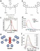
          The potential to image subsurface fluorescent contrast agents at high spatial resolution has facilitated growing interest in short-wave infrared (SWIR) imaging for biomedical applications. The early but growing literature showing improvements in resolution in small animal models suggests this is indeed the case, yet to date, images from larger animal models that more closely recapitulate humans have not been reported. We report the first imaging of SWIR fluorescence in a large animal model. Specifically, we imaged the vascular kinetics of an indocyanine green (ICG) bolus injection during open craniotomy of a mini-pig using a custom SWIR imaging instrument and a clinical-grade surgical microscope that images ICG in the near-infrared-I (NIR-I) window. Fluorescence images in the SWIR were observed to have higher spatial and contrast resolutions throughout the dynamic sequence, particularly in the smallest vessels. Additionally, vessels beneath a surface pool of blood were readily visualized in the SWIR images yet were obscured in the NIR-I channel. These first-in-large-animal observations represent an important translational step and suggest that SWIR imaging may provide higher spatial and contrast resolution images that are robust to the influence of blood.

          Related collections

          Most cited references8

          • Record: found
          • Abstract: found
          • Article: found
          Is Open Access

          Near-Infrared-II (NIR-II) Bioimaging via Off-Peak NIR-I Fluorescence Emission

          Significantly reduced photon scattering and minimal tissue autofluorescence levels in the second biological transparency window (NIR-II; 1000-1700 nm) facilitate higher resolution in vivo biological imaging compared to tradition NIR fluorophores (~700-900 nm). However, the existing palette of NIR-II fluorescent agents including semiconducting inorganic nanomaterials and recently introduced small-molecule organic dyes face significant technical and regulatory hurdles prior to clinical translation. Fortunately, recent spectroscopic characterization of NIR-I dyes (e.g., indocyanine green (ICG), IRDye800CW and IR-12N3) revealed long non-negligible emission tails reaching past 1500 nm. Repurposing the most widely used NIR dye in medicine, in addition to those in the midst of clinical trials creates an accelerated pathway for NIR-II clinical translation. This review focuses on the significant advantage of imaging past 1000 nm with NIR-I fluorophores from both a basic and clinical viewpoint. We further discuss optimizing NIR-I dyes around their NIR-II/shortwave infrared (SWIR) emission, NIR-II emission tail characteristics and prospects of NIR-II imaging with clinically available and commercially available dyes.
            Bookmark
            • Record: found
            • Abstract: found
            • Article: not found

            Review of short-wave infrared spectroscopy and imaging methods for biological tissue characterization.

            We present a review of short-wave infrared (SWIR, defined here as ∼1000 to 2000 nm) spectroscopy and imaging techniques for biological tissue optical property characterization. Studies indicate notable SWIR absorption features of tissue constituents including water (near 1150, 1450, and 1900 nm), lipids (near 1040, 1200, 1400, and 1700 nm), and collagen (near 1200 and 1500 nm) that are much more prominent than corresponding features observed in the visible and near-infrared (VIS-NIR, defined here as ∼400 to 1000 nm). Furthermore, the wavelength dependence of the scattering coefficient has been observed to follow a power-law decay from the VIS-NIR to the SWIR region. Thus, the magnitude of tissue scattering is lower at SWIR wavelengths than that observed at VIS or NIR wavelengths, potentially enabling increased penetration depth of incident light at SWIR wavelengths that are not highly absorbed by the aforementioned chromophores. These aspects of SWIR suggest that the tissue spectroscopy and imaging in this range of wavelengths have the potential to provide enhanced sensitivity (relative to VIS-NIR measurements) to chromophores such as water and lipids, thereby helping to characterize changes in the concentrations of these chromophores due to conditions such as atherosclerotic plaque, breast cancer, and burns.
              Bookmark
              • Record: found
              • Abstract: found
              • Article: not found

              Microscope-integrated near-infrared indocyanine green videoangiography during surgery of intracranial aneurysms: the Helsinki experience.

              Microscope-integrated near-infrared indocyanine green videoangiography (ICG-VA) is a new method of intraoperative blood flow assessment. The objective of this study was to evaluate the reliability of this technique in the evaluation of neck residuals and patency of branches after microneurosurgical clipping of intracranial aneurysms (IAs). During a period of 14 months, between November 2005 and December 2006, 289 patients with intracranial aneurysms were operated on in our institution. Intraoperative ICG-VA was performed during microneurosurgical clipping of 239 IAs in 190 patients. Postoperative computed tomography and computed tomography angiography (CTA) were performed for all patients. Intraoperative interpretation of ICG-VA in assessing the neck residual or the patency of vessels after clipping of each single aneurysm were recorded and correlated with postoperative CTA and/or digital subtraction angiography. Postoperative imaging studies revealed no incomplete occlusions of aneurysm domes. Unexpected neck residuals were observed in 14 aneurysms (6%). There were no parent artery occlusions. Unexpected branch occlusions including both major and minor branching arteries were observed in 15 aneurysms (6%). Indocyanine green videoangiograph is a simple and fast method of blood flow assessment with acceptable reliability. Indocyanine green videoangiograph can provide real-time information to assess blood flow in vessels of different size as well as the occlusion of the aneurysm. Intraoperative assessment of blood flow in the perforating branches is one of the most important advantages. In selected cases such as giant, complex, and deep-sited aneurysms or when the quality of image in ICG-VA is not adequate, other methods of intraoperative blood flow assessment should be considered.
                Bookmark

                Author and article information

                Journal
                J Biomed Opt
                J Biomed Opt
                JBOPFO
                JBO
                Journal of Biomedical Optics
                Society of Photo-Optical Instrumentation Engineers
                1083-3668
                1560-2281
                10 August 2019
                August 2019
                10 August 2019
                : 24
                : 8
                : 080501
                Affiliations
                [a ]Dartmouth College , Thayer School of Engineering, Hanover, New Hampshire, United States
                [b ]Illinois Institute of Technology , Department of Biomedical Engineering, Chicago, Illinois, United States
                [c ]Dartmouth–Hitchcock Medical Center , Department of Surgery, Lebanon, New Hampshire, United States
                Author notes
                [* ]Address all correspondence to Scott C. Davis, E-mail: Scott.C.Davis@ 123456dartmouth.edu
                Author information
                https://orcid.org/0000-0003-1667-3026
                Article
                JBO-190145LR 190145LR
                10.1117/1.JBO.24.8.080501
                6689142
                31401816
                5f0bbcce-6b29-4334-8793-c131a4643ab4
                © The Authors. Published by SPIE under a Creative Commons Attribution 4.0 Unported License. Distribution or reproduction of this work in whole or in part requires full attribution of the original publication, including its DOI.
                History
                : 7 May 2019
                : 15 July 2019
                Page count
                Figures: 4, Tables: 0, References: 13, Pages: 4
                Funding
                Funded by: National Institutes of Health https://doi.org/10.13039/100000002
                Award ID: R01CA184354
                Award ID: R01CA188491
                Award ID: R01CA167413
                Award ID: K99CA190890
                Funded by: Hitchcock Foundation https://doi.org/10.13039/100001170
                Award ID: HF 250-4083
                Categories
                JBO Letters
                Letter
                Custom metadata
                JBO Letters

                Biomedical engineering
                short-wave infrared,near-infrared-ii window,indocyanine green angiography,medical imaging,fluorescence-guided surgery

                Comments

                Comment on this article