15
views
0
recommends
+1 Recommend
0 collections
    0
    shares
      • Record: found
      • Abstract: found
      • Article: found
      Is Open Access

      Modulation of rabbit corneal epithelial cells fate using embryonic stem cell extract

      research-article

      Read this article at

      Bookmark
          There is no author summary for this article yet. Authors can add summaries to their articles on ScienceOpen to make them more accessible to a non-specialist audience.

          Abstract

          Purpose

          To develop a new culture system to cultivate differentiated autologous cells in vitro for cell therapy and tissue engineering.

          Methods

          After incubation in murine embryonic stem cell (ESC) extract for 1 h, streptolysin-O (SLO) permeabilized cells were resealed with CaCl 2 and continually cultured for weeks. The morphological study was analyzed by light microscopy. Isolated colonies were selected and expanded to establish cell lines. Octamer-4 (Oct-4), stage-specific embryonic antigen-1 (SSEA-1), transformation-related protein 63 (p63), ATP-binding cassette subfamily G, member 2 (ABCG2), and cytokeratin3 (K3) were detected by indirect immunofluorescent staining. Oct-4, K3, and p63 were also detected by RT–PCR analysis. To examine the stemness characteristics of the induced cells, both alkaline phosphatase (AKP) staining and tumorigenicity detection were performed, respectively.

          Results

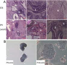
          Reprogramming was induced in corneal epithelial cells. The reprogrammed cells showed characteristics similar to ESCs in the early weeks, including colony formation, positive AKP staining, and multi-potential differentiation in vivo. Oct-4 and SSEA1 protein expression was upregulated. However, these changes were not persistent or stable. With the passage of time, the colonies became flat. The ESC markers were downregulated, while epithelial cell related proteins gradually increased.

          Conclusions

          Less terminal differentiated rabbit corneal epithelial cells could be induced to a more pluripotent state with embryonic stem cell extract (ESC-E). These cells have the potential to return to the beginning of their own lineage and obtain the ability of long-term growth. Our findings indicate that this culture system can generate low-immunogenic autologous cells for use in regenerative medicine.

          Related collections

          Most cited references14

          • Record: found
          • Abstract: found
          • Article: not found

          p63 identifies keratinocyte stem cells.

          The proliferative compartment of stratified squamous epithelia consists of stem and transient amplifying (TA) keratinocytes. Some polypeptides are more abundant in putative epidermal stem cells than in TA cells, but no polypeptide confined to the stem cells has yet been identified. Here we show that the p63 transcription factor, a p53 homologue essential for regenerative proliferation in epithelial development, distinguishes human keratinocyte stem cells from their TA progeny. Within the cornea, nuclear p63 is expressed by the basal cells of the limbal epithelium, but not by TA cells covering the corneal surface. Human keratinocyte stem and TA cells when isolated in culture give rise to holoclones and paraclones, respectively. We show by clonal analysis that p63 is abundantly expressed by epidermal and limbal holoclones, but is undetectable in paraclones. TA keratinocytes, immediately after their withdrawal from the stem cell compartment (meroclones), have greatly reduced p63, even though they possess very appreciable proliferative capacity. Clonal evolution (i.e., generation of TA cells from precursor stem cells) is promoted by the sigma isoform of the 14-3-3 family of proteins. Keratinocytes whose 14-3-3final sigma has been down-regulated remain in the stem cell compartment and maintain p63 during serial cultivation. The identification of p63 as a keratinocyte stem cell marker will be of practical importance for the clinical application of epithelial cultures in cell therapy as well as for studies on epithelial tumorigenesis.
            Bookmark
            • Record: found
            • Abstract: found
            • Article: not found

            ABCG2 transporter identifies a population of clonogenic human limbal epithelial cells.

            ABCG2, a member of the ATP binding cassette (ABC) transporters, has been identified as a molecular determinant for bone marrow stem cells and proposed as a universal marker for stem cells. This study investigates ABCG2 expression and its potential as a marker that identifies human limbal epithelial stem cells. ABCG2 expression was evaluated by immunofluorescent and immunohistochemical staining, laser scanning confocal microscopy, flow cytometry, and semiquantitative reverse transcription-polymerase chain reaction. Cells selected from primary limbal epithelial cultures by flow cytometry with ABCG2 monoclonal antibody (mAb) or Hoechst 33342 dye staining were evaluated for their gene expression and colony-forming efficiency (CFE). ABCG2 protein was mainly located in the basal cells of limbal epithelia but not in the limbal suprabasal and corneal epithelia. ABCG2 staining was also observed in primary limbal epithelial cultures. Limbal epithelia express higher levels of ABCG2 and DeltaNp63 mRNAs than corneal epithelia. Labeling with ABCG2 mAb yielded 2.5%-3.0% positive cells by flow cytometry. The ABCG2-positive cells exhibited greater CFE on a 3T3 fibroblast feeder layer than ABCG2-negative cells. A side population (SP) was detected by the Hoechst 33342 exclusion assay. SP cells displayed stronger expression of ABCG2 and DeltaNp63 mRNA and greater CFE than the non-SP cells. In conclusion, these findings demonstrate that ABCG2 transporter was exclusively expressed by limbal basal cells and that the ABCG2-positive and SP cells possess enriched stem cell properties, suggesting for the first time that ABCG2 could serve as a marker to identify the putative limbal epithelial stem cells.
              Bookmark
              • Record: found
              • Abstract: found
              • Article: not found

              Different intermediate-sized filaments distinguished by immunofluorescence microscopy.

              The major protein of intermediate-sized filaments in mouse 3T3 cells, for which the name vimentin is proposed, has a molecular weight of 57,000. Antibodies against vimentin and antibodies against prekeratin have been used in parallel in immunofluorescence microscopy on a variety of cultured cells as well as on frozen tissue sections. Both antibodies decorate extended wavy arrays of filaments that are different from microfilaments and microtubules. Intermediate filament bundles decorated by antibodies against prekeratin are predominant in many epithelial cells, including epithelia-derived tumor cells, and are not decorated by antibodies to vimentin. In contrast, intermediate filaments decorated by antibodies against vimentin are widespread among nonmuscle cells of mesenchymal origin, including transformed cells, and also occur in other cells. Perinuclear whorls of aggregates of intermediate filaments induced by prolonged treatment with Colcemid generally show strong decoration with antibodies against vimentin. No significant reaction with either antiserum has been observed in muscle structures or in brain nerve tissue. These observations show that intermediate filaments with similar ultrastructure and solubility characteristics can be distinguished immunologically.
                Bookmark

                Author and article information

                Journal
                Mol Vis
                MV
                Molecular Vision
                Molecular Vision
                1090-0535
                2010
                23 June 2010
                : 16
                : 1154-1161
                Affiliations
                [1 ]State Key Laboratory of Ophthalmology, Zhongshan Ophthalmic Center, Sun Yat-sen University, Guangzhou, Guangdong, China
                [2 ]Center for Stem cell Biology and Tissue Engineering, Sun Yat-sen University, Guangzhou, Guangdong, China
                Author notes
                Correspondence to: Zhichong Wang, State Key Laboratory of Ophthalmology, Zhongshan Ophthalmic Center and Center for Stem cell Biology and Tissue Engineering, SunYat-senUniversity, 54 Xian Lie Nan Road, Guangzhou, Guangdong 510060, China; Phone: +86 20 8733 0379; FAX: +86-20-87330389; email: wzc001@ 123456hotmail.com
                Article
                128 2010MOLVIS0169
                2901191
                20664691
                5f0be11d-951a-4228-9990-ebef78fe0fc7
                Copyright © 2010 Molecular Vision.

                This is an open-access article distributed under the terms of the Creative Commons Attribution License, which permits unrestricted use, distribution, and reproduction in any medium, provided the original work is properly cited.

                History
                : 28 April 2010
                : 15 June 2010
                Categories
                Research Article
                Custom metadata
                Export to XML
                Wang

                Vision sciences
                Vision sciences

                Comments

                Comment on this article