21
views
0
recommends
+1 Recommend
0 collections
    0
    shares
      • Record: found
      • Abstract: found
      • Article: found
      Is Open Access

      Altered functional connectivity of amygdala underlying the neuromechanism of migraine pathogenesis

      research-article

      Read this article at

      Bookmark
          There is no author summary for this article yet. Authors can add summaries to their articles on ScienceOpen to make them more accessible to a non-specialist audience.

          Abstract

          Background

          The amygdala is a large grey matter complex in the limbic system, and it may contribute in the neurolimbic pain network in migraine. However, the detailed neuromechanism remained to be elucidated. The objective of this study is to investigate the amygdala structural and functional changes in migraine and to elucidate the mechanism of neurolimbic pain-modulating in the migraine pathogenesis.

          Methods

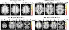
          Conventional MRI, 3D structure images and resting state functional MRI were performed in 18 normal controls (NC), 18 patients with episodic migraine (EM), and 16 patients with chronic migraine (CM). The amygdala volume was measured using FreeSurfer software and the functional connectivity (FC) of bilateral amygdala was computed over the whole brain. Analysis of covariance was performed on the individual FC maps among groups.

          Results

          The increased FC of left amygdala was observed in EM compared with NC, and the decreased of right amygdala was revealed in CM compared with NC. The increased FC of bilateral amygdala was observed in CM compared with EM. The correlation analysis showed a negative correlation between the score of sleep quality (0, normal; 1, mild sleep disturbance; 2, moderate sleep disturbance; 3, serious sleep disturbance) and the increased FC strength of left amygdala in EM compared with NC, and a positive correlation between the score of sleep quality and the increased FC strength of left amygdala in CM compared with EM, and other clinical variables showed no significant correlation with altered FC of amygdala.

          Conclusions

          The altered functional connectivity of amygdala demonstrated that neurolimbic pain network contribute in the EM pathogenesis and CM chronicization.

          Related collections

          Most cited references32

          • Record: found
          • Abstract: found
          • Article: not found

          Functional imaging of brain responses to pain. A review and meta-analysis (2000).

          Brain responses to pain, assessed through positron emission tomography (PET) and functional magnetic resonance imaging (fMRI) are reviewed. Functional activation of brain regions are thought to be reflected by increases in the regional cerebral blood flow (rCBF) in PET studies, and in the blood oxygen level dependent (BOLD) signal in fMRI. rCBF increases to noxious stimuli are almost constantly observed in second somatic (SII) and insular regions, and in the anterior cingulate cortex (ACC), and with slightly less consistency in the contralateral thalamus and the primary somatic area (SI). Activation of the lateral thalamus, SI, SII and insula are thought to be related to the sensory-discriminative aspects of pain processing. SI is activated in roughly half of the studies, and the probability of obtaining SI activation appears related to the total amount of body surface stimulated (spatial summation) and probably also by temporal summation and attention to the stimulus. In a number of studies, the thalamic response was bilateral, probably reflecting generalised arousal in reaction to pain. ACC does not seem to be involved in coding stimulus intensity or location but appears to participate in both the affective and attentional concomitants of pain sensation, as well as in response selection. ACC subdivisions activated by painful stimuli partially overlap those activated in orienting and target detection tasks, but are distinct from those activated in tests involving sustained attention (Stroop, etc.). In addition to ACC, increased blood flow in the posterior parietal and prefrontal cortices is thought to reflect attentional and memory networks activated by noxious stimulation. Less noted but frequent activation concerns motor-related areas such as the striatum, cerebellum and supplementary motor area, as well as regions involved in pain control such as the periaqueductal grey. In patients, chronic spontaneous pain is associated with decreased resting rCBF in contralateral thalamus, which may be reverted by analgesic procedures. Abnormal pain evoked by innocuous stimuli (allodynia) has been associated with amplification of the thalamic, insular and SII responses, concomitant to a paradoxical CBF decrease in ACC. It is argued that imaging studies of allodynia should be encouraged in order to understand central reorganisations leading to abnormal cortical pain processing. A number of brain areas activated by acute pain, particularly the thalamus and anterior cingulate, also show increases in rCBF during analgesic procedures. Taken together, these data suggest that hemodynamic responses to pain reflect simultaneously the sensory, cognitive and affective dimensions of pain, and that the same structure may both respond to pain and participate in pain control. The precise biochemical nature of these mechanisms remains to be investigated.
            Bookmark
            • Record: found
            • Abstract: found
            • Article: not found

            Altered functional magnetic resonance imaging resting-state connectivity in periaqueductal gray networks in migraine.

            The periaqueductal gray matter (PAG), a known modulator of somatic pain transmission, shows evidence of interictal functional and structural abnormalities in migraineurs, which may contribute to hyperexcitability along spinal and trigeminal nociceptive pathways, and lead to the migraine attack. The aim of this study was to examine functional connectivity of the PAG in migraine. Using resting-state functional MRI, we compared functional connectivity between PAG and a subset of brain areas involved in nociceptive/somatosensory processing and pain modulation in 17 subjects with migraine, during a pain-free state, versus 17 gender- and age-matched controls. We also assessed the relation between intrinsic resting-state correlations within PAG networks and the average monthly frequency of migraine attacks, as well as allodynia. Our findings show stronger connectivity between the PAG and several brain areas within nociceptive and somatosensory processing pathways in migraineurs versus controls. In addition, as the monthly frequency of migraine attacks worsens, the strength of the connectivity in some areas within these pathways increases, whereas a significant decrease in functional resting-state connectivity between the PAG and brain regions with a predominant role in pain modulation (prefrontal cortex, anterior cingulate, amygdala) can be evidenced. Finally, migraineurs with a history of allodynia exhibit significantly reduced connectivity between PAG, prefrontal regions, and anterior cingulate compared to migraineurs without allodynia. These data reveal interictal dysfunctional dynamics within pain pathways in migraine manifested as an impairment of the descending pain modulatory circuits, likely leading to loss of pain inhibition, and hyperexcitability primarily in nociceptive areas. Copyright © 2011 American Neurological Association.
              Bookmark
              • Record: found
              • Abstract: found
              • Article: not found

              Headache and sleep: examination of sleep patterns and complaints in a large clinical sample of migraineurs.

              This study characterized sleep parameters and complaints in a large clinical sample of migraineurs and examined sleep complaints in relation to headache frequency and severity. The relationship between headache and sleep has been documented at least anecdotally in medical literature for well over a century and clinical texts allude to the importance of sleep as a headache precipitant. A small number of empirical studies have emerged, but the precise nature and magnitude of the headache/sleep association and underlying mechanisms remain poorly understood. In this investigation, 1283 migraineurs were drawn from 1480 consecutive headache sufferers presenting for evaluation to a tertiary headache clinic. Patients underwent a physical examination and structured interview assessing a variety of sleep, headache, and demographic variables. Migraine was diagnosed according the IHS criteria (1.1 to 1.6 diagnostic codes). Migraineurs were 84% female, with a mean age of 37.4 years. Groups were formed based on patient's average nocturnal sleep patterns, including short, normal, and long sleep groups, and were compared on headache variables. Sleep complaints were common and associated with headache in a sizeable proportion of patients. Over half of migraineurs reported difficulty initiating and maintaining sleep at least occasionally. Many in this sample reported chronically shortened sleep patterns similar to that observed in persons with insomnia, with 38% of patients sleeping on average 6 hours per night. Migraines were triggered by sleep disturbance in 50% of patients. "Awakening headaches" or headaches awakening them from sleep were reported by 71% of patients. Interestingly, sleep was also a common palliative agent for headache; 85% of migraineurs indicated that they chose to sleep or rest because of headache and 75% were forced to sleep or rest because of headache. Patients with chronic migraine reported shorter nightly sleep times than those with episodic migraine, and were more likely to exhibit trouble falling asleep, staying asleep, sleep triggering headache, and choosing to sleep because of headache. Short sleepers (ie, average sleep period 6 hours) exhibited significantly more frequent and more severe headaches than individuals who slept longer and were more likely to exhibit morning headaches on awakening. These data support earlier research and anecdotal observations of a substantial sleep/migraine relationship, and implicate sleep disturbance in specific headache patterns and severity. The short sleep group, who routinely slept 6 hours per night, exhibited the more severe headache patterns and more sleep-related headache. Sleep complaints occurred with greater frequency among chronic than episodic migraineurs. Future research may identify possible mediating factors such as primary sleep and mood disorders. Prospective studies are needed to determine if normalizing sleep times in the short sleeps would impact headache threshold.
                Bookmark

                Author and article information

                Contributors
                cjr.malin@vip.163.com
                yusy1963@126.com
                Journal
                J Headache Pain
                J Headache Pain
                The Journal of Headache and Pain
                Springer Milan (Milan )
                1129-2369
                1129-2377
                23 January 2017
                23 January 2017
                2017
                : 18
                : 1
                : 7
                Affiliations
                [1 ]ISNI 0000 0004 1761 8894, GRID grid.414252.4, Department of Radiology, , Chinese PLA General Hospital, ; 28 Fuxing Road, Beijing, 100853 China
                [2 ]ISNI 0000 0004 1761 8894, GRID grid.414252.4, Department of Neurology, , Chinese PLA General Hospital, ; 28 Fuxing Road, Beijing, 100853 China
                [3 ]GRID grid.452517.0, Department of Radiology, , Hainan Branch of Chinese PLA General Hospital, ; Beijing, 100853 China
                Article
                722
                10.1186/s10194-017-0722-5
                5256627
                28116559
                60df548a-1c16-4344-8525-001911130895
                © The Author(s). 2017

                Open AccessThis article is distributed under the terms of the Creative Commons Attribution 4.0 International License ( http://creativecommons.org/licenses/by/4.0/), which permits unrestricted use, distribution, and reproduction in any medium, provided you give appropriate credit to the original author(s) and the source, provide a link to the Creative Commons license, and indicate if changes were made.

                History
                : 20 November 2016
                : 2 January 2017
                Funding
                Funded by: the National Natural Sciences Foundation of China
                Award ID: 81371514
                Award Recipient :
                Funded by: the Special Financial Grant from the China Postdoctoral Science Foundation
                Award ID: 2014T70960
                Award Recipient :
                Categories
                Research Article
                Custom metadata
                © The Author(s) 2017

                Anesthesiology & Pain management
                migraine,amygdala,neurolimbic pain network,functional connectivity,fmri

                Comments

                Comment on this article