36
views
0
recommends
+1 Recommend
0 collections
    0
    shares
      • Record: found
      • Abstract: found
      • Article: found
      Is Open Access

      Automated segmentation by pixel classification of retinal layers in ophthalmic OCT images

      research-article

      Read this article at

      Bookmark
          There is no author summary for this article yet. Authors can add summaries to their articles on ScienceOpen to make them more accessible to a non-specialist audience.

          Abstract

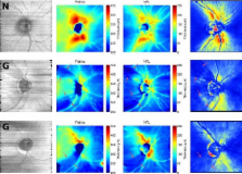
          Current OCT devices provide three-dimensional (3D) in-vivo images of the human retina. The resulting very large data sets are difficult to manually assess. Automated segmentation is required to automatically process the data and produce images that are clinically useful and easy to interpret. In this paper, we present a method to segment the retinal layers in these images. Instead of using complex heuristics to define each layer, simple features are defined and machine learning classifiers are trained based on manually labeled examples. When applied to new data, these classifiers produce labels for every pixel. After regularization of the 3D labeled volume to produce a surface, this results in consistent, three-dimensionally segmented layers that match known retinal morphology. Six labels were defined, corresponding to the following layers: Vitreous, retinal nerve fiber layer (RNFL), ganglion cell layer & inner plexiform layer, inner nuclear layer & outer plexiform layer, photoreceptors & retinal pigment epithelium and choroid. For both normal and glaucomatous eyes that were imaged with a Spectralis (Heidelberg Engineering) OCT system, the five resulting interfaces were compared between automatic and manual segmentation. RMS errors for the top and bottom of the retina were between 4 and 6 μm, while the errors for intra-retinal interfaces were between 6 and 15 μm. The resulting total retinal thickness maps corresponded with known retinal morphology. RNFL thickness maps were compared to GDx (Carl Zeiss Meditec) thickness maps. Both maps were mostly consistent but local defects were better visualized in OCT-derived thickness maps.

          Related collections

          Most cited references16

          • Record: found
          • Abstract: not found
          • Article: not found

          Support-vector networks

            Bookmark
            • Record: found
            • Abstract: found
            • Article: found
            Is Open Access

            Automatic segmentation of seven retinal layers in SDOCT images congruent with expert manual segmentation

            Segmentation of anatomical and pathological structures in ophthalmic images is crucial for the diagnosis and study of ocular diseases. However, manual segmentation is often a time-consuming and subjective process. This paper presents an automatic approach for segmenting retinal layers in Spectral Domain Optical Coherence Tomography images using graph theory and dynamic programming. Results show that this method accurately segments eight retinal layer boundaries in normal adult eyes more closely to an expert grader as compared to a second expert grader.
              Bookmark
              • Record: found
              • Abstract: found
              • Article: not found

              Automated 3-D intraretinal layer segmentation of macular spectral-domain optical coherence tomography images.

              With the introduction of spectral-domain optical coherence tomography (OCT), much larger image datasets are routinely acquired compared to what was possible using the previous generation of time-domain OCT. Thus, the need for 3-D segmentation methods for processing such data is becoming increasingly important. We report a graph-theoretic segmentation method for the simultaneous segmentation of multiple 3-D surfaces that is guaranteed to be optimal with respect to the cost function and that is directly applicable to the segmentation of 3-D spectral OCT image data. We present two extensions to the general layered graph segmentation method: the ability to incorporate varying feasibility constraints and the ability to incorporate true regional information. Appropriate feasibility constraints and cost functions were learned from a training set of 13 spectral-domain OCT images from 13 subjects. After training, our approach was tested on a test set of 28 images from 14 subjects. An overall mean unsigned border positioning error of 5.69+/-2.41 microm was achieved when segmenting seven surfaces (six layers) and using the average of the manual tracings of two ophthalmologists as the reference standard. This result is very comparable to the measured interobserver variability of 5.71+/-1.98 microm.
                Bookmark

                Author and article information

                Journal
                Biomed Opt Express
                BOE
                Biomedical Optics Express
                Optical Society of America
                2156-7085
                27 May 2011
                1 June 2011
                27 May 2011
                : 2
                : 6
                : 1743-1756
                Affiliations
                [1 ]Rotterdam Ophthalmic Institute, Rotterdam Eye Hospital, P.O. Box 70030, 3000 LM Rotterdam, The Netherlands
                [2 ]Glaucoma Service, Rotterdam Eye Hospital, P.O. Box 70030, 3000 LM Rotterdam, The Netherlands
                [3 ]Dept. of Physics and Astronomy, VU University, De Boelelaan 1081, 1081 HV Amsterdam, The Netherlands
                [4 ]LaserLaB Amsterdam, VU University, De Boelelaan 1081, 1081 HV Amsterdam, The Netherlands
                Author notes
                Article
                144515
                10.1364/BOE.2.001743
                3114239
                21698034
                6197b2ff-0a97-4f43-a5cf-ca2acda03b4b
                ©2011 Optical Society of America

                This is an open-access article distributed under the terms of the Creative Commons Attribution-Noncommercial-No Derivative Works 3.0 Unported License, which permits download and redistribution, provided that the original work is properly cited. This license restricts the article from being modified or used commercially.

                History
                : 27 March 2011
                : 13 May 2011
                : 20 May 2011
                Funding
                Funded by: The Rotterdam Eye Hospital Research Foundation, Rotterdam, The Netherlands
                Funded by: Stichting Oogfonds Nederland, Utrecht, The Netherlands
                Funded by: Glaucoomfonds, Utrecht, The Netherlands
                Funded by: Landelijke Stichting voor Blinden en Slechtzienden, Utrecht, The Netherlands
                Funded by: Stichting voor Ooglijders, Rotterdam, The Netherlands
                Funded by: Stichting Nederlands Oogheelkundig Onderzoek, Nijmegen, The Netherlands
                Categories
                Image Processing
                Custom metadata
                True
                12

                Vision sciences
                (100.5010) pattern recognition,(100.2960) image analysis,(170.4580) optical diagnostics for medicine,(170.4500) optical coherence tomography,(100.0100) image processing,(170.4470) ophthalmology

                Comments

                Comment on this article