31
views
0
recommends
+1 Recommend
0 collections
    0
    shares
      • Record: found
      • Abstract: found
      • Article: found
      Is Open Access

      Amygdala Plasticity and Pain

      review-article
      1 , 1 , 2 ,
      Pain Research & Management
      Hindawi

      Read this article at

      Bookmark
          There is no author summary for this article yet. Authors can add summaries to their articles on ScienceOpen to make them more accessible to a non-specialist audience.

          Abstract

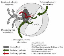
          The amygdala is a limbic brain region that plays a key role in emotional processing, neuropsychiatric disorders, and the emotional-affective dimension of pain. Preclinical and clinical studies have identified amygdala hyperactivity as well as impairment of cortical control mechanisms in pain states. Hyperactivity of basolateral amygdala (BLA) neurons generates enhanced feedforward inhibition and deactivation of the medial prefrontal cortex (mPFC), resulting in pain-related cognitive deficits. The mPFC sends excitatory projections to GABAergic neurons in the intercalated cell mass (ITC) in the amygdala, which project to the laterocapsular division of the central nucleus of the amygdala (CeLC; output nucleus) and serve gating functions for amygdala output. Impairment of these cortical control mechanisms allows the development of amygdala pain plasticity. Mechanisms of abnormal amygdala activity in pain with particular focus on loss of cortical control mechanisms as well as new strategies to correct pain-related amygdala dysfunction will be discussed in the present review.

          Related collections

          Most cited references154

          • Record: found
          • Abstract: found
          • Article: not found

          The amygdala modulates the consolidation of memories of emotionally arousing experiences.

          Converging findings of animal and human studies provide compelling evidence that the amygdala is critically involved in enabling us to acquire and retain lasting memories of emotional experiences. This review focuses primarily on the findings of research investigating the role of the amygdala in modulating the consolidation of long-term memories. Considerable evidence from animal studies investigating the effects of posttraining systemic or intra-amygdala infusions of hormones and drugs, as well as selective lesions of specific amygdala nuclei, indicates that (a) the amygdala mediates the memory-modulating effects of adrenal stress hormones and several classes of neurotransmitters; (b) the effects are selectively mediated by the basolateral complex of the amygdala (BLA); (c) the influences involve interactions of several neuromodulatory systems within the BLA that converge in influencing noradrenergic and muscarinic cholinergic activation; (d) the BLA modulates memory consolidation via efferents to other brain regions, including the caudate nucleus, nucleus accumbens, and cortex; and (e) the BLA modulates the consolidation of memory of many different kinds of information. The findings of human brain imaging studies are consistent with those of animal studies in suggesting that activation of the amygdala influences the consolidation of long-term memory; the degree of activation of the amygdala by emotional arousal during encoding of emotionally arousing material (either pleasant or unpleasant) correlates highly with subsequent recall. The activation of neuromodulatory systems affecting the BLA and its projections to other brain regions involved in processing different kinds of information plays a key role in enabling emotionally significant experiences to be well remembered.
            Bookmark
            • Record: found
            • Abstract: found
            • Article: not found

            Dissociable roles of prelimbic and infralimbic cortices, ventral hippocampus, and basolateral amygdala in the expression and extinction of conditioned fear.

            Current models of conditioned fear expression and extinction involve the basolateral amygdala (BLA), ventral medial prefrontal cortex (vmPFC), and the hippocampus (HPC). There is some disagreement with respect to the specific roles of these structures, perhaps due to subregional differences within each area. For example, growing evidence suggests that infralimbic (IL) and prelimbic (PL) subregions of vmPFC have opposite influences on fear expression. Moreover, it is the ventral HPC (vHPC), rather than the dorsal HPC, that projects to vmPFC and BLA. To help determine regional specificity, we used small doses of the GABA(A) agonist muscimol to selectively inactivate IL, PL, BLA, or vHPC in an auditory fear conditioning and extinction paradigm. Infusions were performed prior to extinction training, allowing us to assess the effects on both fear expression and subsequent extinction memory. Inactivation of IL had no effect on fear expression, but impaired the within-session acquisition of extinction as well as extinction memory. In contrast, inactivation of PL impaired fear expression, but had no effect on extinction memory. Inactivation of the BLA or vHPC impaired both fear expression and extinction memory. Post-extinction inactivations had no effect in any structure. We suggest a model in which amygdala-dependent fear expression is modulated by inputs from PL and vHPC, whereas extinction memory requires extinction-induced plasticity in IL, BLA, and/or vHPC.
              Bookmark
              • Record: found
              • Abstract: found
              • Article: not found

              Chronic back pain is associated with decreased prefrontal and thalamic gray matter density.

              The role of the brain in chronic pain conditions remains speculative. We compared brain morphology of 26 chronic back pain (CBP) patients to matched control subjects, using magnetic resonance imaging brain scan data and automated analysis techniques. CBP patients were divided into neuropathic, exhibiting pain because of sciatic nerve damage, and non-neuropathic groups. Pain-related characteristics were correlated to morphometric measures. Neocortical gray matter volume was compared after skull normalization. Patients with CBP showed 5-11% less neocortical gray matter volume than control subjects. The magnitude of this decrease is equivalent to the gray matter volume lost in 10-20 years of normal aging. The decreased volume was related to pain duration, indicating a 1.3 cm3 loss of gray matter for every year of chronic pain. Regional gray matter density in 17 CBP patients was compared with matched controls using voxel-based morphometry and nonparametric statistics. Gray matter density was reduced in bilateral dorsolateral prefrontal cortex and right thalamus and was strongly related to pain characteristics in a pattern distinct for neuropathic and non-neuropathic CBP. Our results imply that CBP is accompanied by brain atrophy and suggest that the pathophysiology of chronic pain includes thalamocortical processes.
                Bookmark

                Author and article information

                Contributors
                Journal
                Pain Res Manag
                Pain Res Manag
                PRM
                Pain Research & Management
                Hindawi
                1203-6765
                1918-1523
                2017
                10 December 2017
                : 2017
                : 8296501
                Affiliations
                1Department of Pharmacology and Neuroscience, Texas Tech University Health Sciences Center, School of Medicine, Lubbock, TX, USA
                2Center of Excellence for Translational Neuroscience and Therapeutics, Texas Tech University Health Sciences Center, Lubbock, TX, USA
                Author notes

                Academic Editor: Susanne Becker

                Author information
                http://orcid.org/0000-0001-6158-8635
                Article
                10.1155/2017/8296501
                5742506
                29302197
                653adf5f-90ec-4cc9-8fe5-f5777eeaf992
                Copyright © 2017 Jeremy M. Thompson and Volker Neugebauer.

                This is an open access article distributed under the Creative Commons Attribution License, which permits unrestricted use, distribution, and reproduction in any medium, provided the original work is properly cited.

                History
                : 20 July 2017
                : 2 November 2017
                Funding
                Funded by: National Institutes of Health
                Award ID: NS081121
                Award ID: NS038261
                Categories
                Review Article

                Comments

                Comment on this article