5
views
0
recommends
+1 Recommend
0 collections
    0
    shares
      • Record: found
      • Abstract: found
      • Article: found
      Is Open Access

      Chiasmal herniation following treatment of pituitary macroadenoma

      research-article

      Read this article at

      Bookmark
          There is no author summary for this article yet. Authors can add summaries to their articles on ScienceOpen to make them more accessible to a non-specialist audience.

          Abstract

          Purpose

          To evaluate whether the occurrence of chiasmal herniation coincides with visual field (VF) deterioration and to compare the course of VF defects in patients with and without radiological chiasmal herniation following treatment of pituitary adenoma.

          Methods

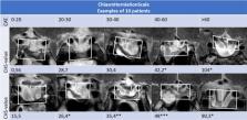
          This retrospective cohort study included 48 pituitary macroadenoma patients with chiasm compression, divided into three groups: Group 1 (N = 12), downward displaced optic chiasm and deteriorated VFs; Group 2 (N = 16), downward displaced optic chiasm; Group 3 (N = 20), control-group matched for tumour size and follow-up VFs, in mean deviation (dB). VFs were compared over time and a severity index, Chiasm Herniation Scale (CHS), for herniation based on radiological parameters was designed.

          Results

          After treatment, all groups showed improvement of VFs (Gr1: 2.97 dB p = 0.097, Gr2: 4.52 dB p = 0.001 and Gr3: 5.16 dB p = 0.000), followed by long-term gradual deterioration. The course of VFs between patients with and without herniation was not significantly different (p = 0.143), neither was there a difference in the course before and after herniation (p = 0.297). The median time till onset of herniation was 40 months (IQR 6 month-10 years) and did not significantly differ (p = 0.172) between the groups. There was no relation between VFs and the degree of herniation (p = 0.729).

          Conclusion

          Herniation does not appear to have clinical relevance with respect to VF outcome. The newly designed CHS is the first scoring system to quantify the severity of herniation and, in the absence of alternatives, may be useful to describe MRI findings to serve future added value in larger sized outcome studies.

          Related collections

          Most cited references41

          • Record: found
          • Abstract: found
          • Article: not found

          Segments of the internal carotid artery: a new classification.

          This study proposes an anatomically based nomenclature for the internal carotid artery (ICA) that can be applied by all disciplines. In 1938, Fischer published a seminal paper describing five segments of the ICA that were designated C1 through C5. These segments were based on the angiographic course of the intracranial ICA rather than its arterial branches or anatomic compartments. Subsequent attempts to apply modern nomenclature to these numerical segments failed to recognize Fischer's original intent of describing patterns of arterial displacement by tumors and, therefore, resulted in a nomenclature that was anatomically inaccurate. Fischer's system was further limited, because segments were numbered opposite the direction of blood flow and the extracranial ICA was excluded. The authors propose a new classification, which includes the entire ICA, uses a numerical scale in the direction of blood flow, and describes the segments of the ICA according to a detailed understanding of the anatomy surrounding the ICA and the compartments through which it travels. Twenty cadaveric specimens with intravascular injection of silicone rubber were used for microscopic dissection and 20 dry skulls were inspected. Histological sections in critical areas were examined. The authors' classification has the following seven segments: C1, cervical; C2, petrous; C3, lacerum; C4 cavernous; C5, clinoid; C6, ophthalmic; and C7, communicating. This classification is practical, accounts for new anatomic information and clinical interests, and clarifies all segments of the ICA.
            Bookmark
            • Record: found
            • Abstract: not found
            • Article: not found

            Health as a Theoretical Concept

              Bookmark
              • Record: found
              • Abstract: found
              • Article: not found

              Diagnosis, treatment, and outcome of pituitary tumors and other abnormal intrasellar masses. Retrospective analysis of 353 patients.

              We reviewed the clinical features, essential laboratory data, pituitary imaging findings (computerized tomography and magnetic resonance imaging), management, and outcome of 353 consecutive patients with the presumptive diagnosis of pituitary tumor investigated from January 1984 through December 1997 at University Hospital, Lausanne, Switzerland. In 18 cases primary empty sella turcica was diagnosed, and in 13 cases of pseudacromegaly there were no endocrine abnormalities. The remaining 322 patients disclosed abnormal pituitary masses, including 275 pituitary adenomas, 18 craniopharyngiomas, 6 cases of primary pituitary hyperplasia, 6 intrasellar meningiomas, 6 cases of distant metastases, 4 intrasellar cysts, 2 chordomas, 1 primary lymphoma, and 1 astrocytoma. Biologic data and immunohistochemical analysis of the excised tissues demonstrated that prolactinomas and nonsecreting adenomas (NSAs) were the most frequent pituitary tumors (40% and 39%, respectively), followed by somatotropic adenomas with acromegaly (11%) and Cushing disease (6%). In contrast with the vast majority of NSAs, which significantly expressed glycoprotein hormones in tissue without secreting them, there was a small group of glycoprotein hormone-secreting adenomas (2%), which had a more severe clinical course after surgery. Thirty-eight pituitary masses were incidentally discovered, most of them NSAs. The expansion of pituitary adenomas into the right cavernous sinus was twice as frequent as to the left cavernous sinus. For the differential diagnosis of hyperprolactinemia, basal prolactin (PRL) levels above 85 micrograms/L, in the absence of renal failure and PRL-enhancing drugs, and a PRL increment of less than 30% after thyrotropin-releasing hormone (TRH) accurately ruled out functional hyperprolactinemia due to NSA, and were typical of prolactinomas. For screening and follow-up of acromegaly, basal growth hormone (GH) and insulin-like growth factor 1 (IGF-1) levels, as well as the paradoxical GH response to TRH (present in 2/3 acromegalic patients), could be used as convenient tools, but the most accurate test for diagnosis and prediction of outcome after therapy was GH (lack of) suppression during oral glucose tolerance test. In Cushing disease, single evening plasma cortisol was as good as the overnight dexamethasone suppression test for screening, and a combined dexamethasoneovine corticotropin-releasing hormone (oCRH) test was as accurate as the long dexamethasone suppression test to confirm the diagnosis. Bilateral inferior petrosal sinus catheterization coupled with oCRH test confirmed the pituitary origin of excess adrenocorticotropic hormone (ACTH) in all patients, including those with normal pituitary on magnetic resonance imaging (50% of the cases). However, this procedure failed to predict tumor localization correctly within the pituitary in 21% of patients. Pituitary cysts, meningiomas, and craniopharyngiomas with an intrasellar component were correctly diagnosed based on pituitary imaging in 75%, 67%, and 44% of cases, respectively. The remainder, as well as the cases of pituitary hyperplasia, metastases, and other less frequent pathologies, were initially diagnosed as NSAs or as masses of unknown nature. When surgery was indicated, pituitary adenomas and other intrasellar masses were operated on by the transsphenoidal route, with the exception of 100% of meningiomas, 83% of craniopharyngiomas, and 10% of NSAs, which were operated on by the transcranial route. Favorable late surgical outcome of prolactinomas could be predicted by a restored PRL response to TRH. However, dopamine agonist (DA) therapy, usually resulting in satisfactory control of PRL levels and in tumor shrinkage, progressively displaced surgery as primary treatment for prolactinomas throughout the study period. After full-term pregnancy, the size of prolactinoma decreased in 7 of 9 patients, and PRL was normal in 2. Surgery was the first treatment for NSAs, with a tumor rela
                Bookmark

                Author and article information

                Contributors
                marjoleintabak@live.nl , mtabak@lumc.nl
                i.c.notting@lumc.nl
                Journal
                Pituitary
                Pituitary
                Pituitary
                Springer US (New York )
                1386-341X
                1573-7403
                15 October 2020
                15 October 2020
                2021
                : 24
                : 1
                : 68-78
                Affiliations
                [1 ]GRID grid.10419.3d, ISNI 0000000089452978, Department of Ophthalmology, , Leiden University Medical Center, ; Leiden, The Netherlands
                [2 ]GRID grid.10419.3d, ISNI 0000000089452978, Department of Medicine, Division of Endocrinology, , Leiden University Medical Center, ; Leiden, The Netherlands
                [3 ]GRID grid.10419.3d, ISNI 0000000089452978, Department of Radiology, , Leiden University Medical Center, ; Leiden, The Netherlands
                [4 ]GRID grid.10419.3d, ISNI 0000000089452978, Department of Neurosurgery, , Leiden University Medical Center, ; Leiden, The Netherlands
                [5 ]GRID grid.10419.3d, ISNI 0000000089452978, Center for Pituitary Tumours Leiden, , Leiden University Medical Center, ; Leiden, The Netherlands
                Author information
                http://orcid.org/0000-0001-6471-7084
                Article
                1088
                10.1007/s11102-020-01088-2
                7864822
                33057947
                66e34242-17c8-46fc-81ff-de87ec7e08fc
                © The Author(s) 2020

                Open AccessThis article is licensed under a Creative Commons Attribution 4.0 International License, which permits use, sharing, adaptation, distribution and reproduction in any medium or format, as long as you give appropriate credit to the original author(s) and the source, provide a link to the Creative Commons licence, and indicate if changes were made. The images or other third party material in this article are included in the article's Creative Commons licence, unless indicated otherwise in a credit line to the material. If material is not included in the article's Creative Commons licence and your intended use is not permitted by statutory regulation or exceeds the permitted use, you will need to obtain permission directly from the copyright holder. To view a copy of this licence, visit http://creativecommons.org/licenses/by/4.0/.

                History
                : 26 September 2020
                Funding
                Funded by: Leiden University Medical Center (LUMC)
                Categories
                Article
                Custom metadata
                © Springer Science+Business Media, LLC, part of Springer Nature 2021

                Medicine
                pituitary adenoma,optic chiasm,chiasmal herniation,visual field,visual deterioration,chiasmapexy

                Comments

                Comment on this article