29
views
0
recommends
+1 Recommend
0 collections
    0
    shares
      • Record: found
      • Abstract: found
      • Article: not found

      Porcine Circovirus Type 2 and Porcine Circovirus‐Associated Disease

      review-article

      Read this article at

      ScienceOpenPublisherPMC
      Bookmark
          There is no author summary for this article yet. Authors can add summaries to their articles on ScienceOpen to make them more accessible to a non-specialist audience.

          Abstract

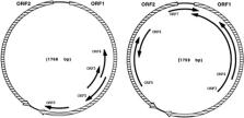
          Porcine circovirus type 2 (PCV2) belongs to the viral family Circoviridae and to the genus Circovirus. Circoviruses are small, single‐stranded nonenveloped DNA viruses that have an unsegmented circular genome. PCV2 is the primary causative agent of several syndromes collectively known as porcine circovirus‐associated disease (PCVAD). Many of the syndromes associated with PCVAD are a result of coinfection with PCV2 virus and other agents such as Mycoplasma and porcine reproductive and respiratory syndrome virus. PCV2 infection is present in every major swine‐producing country in the world, and the number of identified cases of PCVAD is rapidly increasing. In the United States, the disease has cost producers an average of 3–4 dollars per pig with peak losses ranging up to 20 dollars per pig. The importance of this disease has stimulated investigations aimed at identifying risk factors associated with infection and minimizing these risks through modified management practices and development of vaccination strategies. This paper provides an overview of current knowledge relating to PCV2 and PCVAD with an emphasis on information relevant to the swine veterinarian.

          Related collections

          Most cited references135

          • Record: found
          • Abstract: found
          • Article: not found

          A novel DNA virus (TTV) associated with elevated transaminase levels in posttransfusion hepatitis of unknown etiology.

          By means of representational difference analysis, a viral clone (N22) of 500 nucleotides was isolated from serum of a patient (TT) with posttransfusion hepatitis of unknown etiology. The N22 clone showed a poor homology to any reported sequences. Oligonucleotide primers were deduced from the N22 sequence for detecting it by polymerase chain reaction. N22 sequence in serum banded at a sucrose density of 1.26 g/cm3, indicating its association with a viral particle which was designated TT virus (TTV). Since nucleic acids of TTV were sensitive to DNase I, it would be a DNA virus. TTV DNA was detected in sera from three of the five patients with posttransfusion non-A to G hepatitis, including the index case (TT). TTV DNA titers closely correlated with aminotransferase levels in the three patients. These results indicate that TTV would be a novel DNA virus with a possible capacity to induce posttransfusion non-A to G hepatitis. Copyright 1997 Academic Press.
            Bookmark
            • Record: found
            • Abstract: found
            • Article: not found

            Experimental reproduction of postweaning multisystemic wasting syndrome in pigs by dual infection with Mycoplasma hyopneumoniae and porcine circovirus type 2.

            The objectives of this study were to investigate the interactions between Mycoplasma hyopneumoniae and porcine circovirus type 2 (PCV2) and to establish a model for studying the pathogenesis of and testing intervention strategies for the control of PCV2-associated porcine respiratory disease complex (PRDC). Sixty-seven pigs were randomly assigned to four groups. Group 1 (n=17) pigs served as controls, group 2 (n=17) pigs were inoculated with M. hyopneumoniae, group 3 (n=17) pigs were dual infected with M. hyopneumoniae and PCV2, and group 4 (n=16) pigs were inoculated with PCV2. Pigs were inoculated intratracheally with M. hyopneumoniae at 4 weeks of age followed by intranasal inoculation with PCV2 at 6 weeks of age. Dual-infected pigs had moderate dyspnea, lethargy, and reduced weight gain. The overall severity of macroscopic lung lesions, PCV2-associated microscopic lesions in lung and lymphoid tissues, and the amount of PCV2-antigen associated with these lesions were significantly (P <0.05) higher in dual-infected pigs compared with all other groups. Four of 17 (23.5%) dual-infected pigs had decreased growth rate and severe lymphoid depletion and granulomatous lymphadenitis associated with high amounts of PCV2-antigen consistent with postweaning multisystemic wasting syndrome (PMWS). PCV2-antigen in lung tissue was most often associated with M. hyopneumoniae-induced peribronchial lymphoid hyperplasia, suggesting that this is an important site for PCV2 replication in the lung. This study indicates that M. hyopneumoniae potentiates the severity of PCV2-associated lung and lymphoid lesions, increases the amount and prolongs the presence of PCV2-antigen, and increases the incidence of PMWS in pigs.
              Bookmark
              • Record: found
              • Abstract: found
              • Article: not found

              Experimental reproduction of severe disease in CD/CD pigs concurrently infected with type 2 porcine circovirus and porcine reproductive and respiratory syndrome virus.

              Three-week-old cesarean-derived colostrum-deprived (CD/CD) pigs were inoculated with porcine circovirus type 2 (PCV2, n = 19), porcine reproductive and respiratory syndrome virus (PRRSV, n = 13), concurrent PCV2 and PRRSV (PCV2/PRRSV, n = 17), or a sham inoculum (n = 12) to compare the independent and combined effects of these agents. Necropsies were performed at 7, 10, 14, 21, 35, and 49 days postinoculation (dpi) or when pigs became moribund. By 10 dpi, PCV2/PRRSV-inoculated pigs had severe dyspnea, lethargy, and occasional icterus; after 10 dpi, mortality in this group was 10/11 (91%), and all PCV2/ PRRSV-inoculated pigs were dead by 20 dpi. PCV2-inoculated pigs developed lethargy and sporadic icterus, and 8/19 (42%) developed exudative epidermitis; mortality was 5/19 (26%). PRRSV-inoculated pigs developed dyspnea and mild lethargy that resolved by 28 dpi. Microscopic lesions consistent with postweaning multisystemic wasting syndrome (PMWS) were present in both PCV2- and PCV2/PRRSV-inoculated pigs and included lymphoid depletion, necrotizing hepatitis, mild necrotizing bronchiolitis, and infiltrates of macrophages that occasionally contained basophilic intracytoplasmic inclusion bodies in lymphoid and other tissues. PCV2/ PRRSV-inoculated pigs also had severe proliferative interstitial pneumonia and more consistent hepatic lesions. The most severe lesions contained the greatest number of PCV2 antigen-containing cells. PRRSV-inoculated pigs had moderate proliferative interstitial pneumonia but did not develop bronchiolar or hepatic lesions or lymphoid depletion. All groups remained seronegative to porcine parvovirus. The results indicate that 1) PCV2 coinfection increases the severity of PRRSV-induced interstitial pneumonia in CD/CD pigs and 2) PCV2 but not PRRSV induces the lymphoid depletion, granulomatous inflammation, and necrotizing hepatitis characteristic of PMWS.
                Bookmark

                Author and article information

                Journal
                J Vet Intern Med
                J. Vet. Intern. Med
                10.1111/(ISSN)1939-1676
                JVIM
                Journal of Veterinary Internal Medicine
                Blackwell Publishing Inc (Malden, USA )
                0891-6640
                1939-1676
                22 September 2009
                Nov-Dec 2009
                : 23
                : 6 ( doiID: 10.1111/jvim.2009.23.issue-6 )
                : 1151-1163
                Affiliations
                [ 1 ]Department of Large Animal Clinical Sciences, Virginia‐Maryland Regional College of Veterinary Medicine, Virginia Polytechnic Institute and State University, Blacksburg, VA
                Author notes
                [*]Corresponding author: Virginia Buechner‐Maxwell, DVM, MS, Diplomate ACVIM, Professor, Virginia Polytechnic Institute and State University, Duck Pond Drive, Blacksburg, VA 24061‐0342; e‐mail: bmax@ 123456vt.edu .
                Article
                JVIM389 389
                10.1111/j.1939-1676.2009.0389.x
                7166794
                19780932
                66e99269-e6d9-4665-bbca-201c8286db66
                Copyright © 2009 by the American College of Veterinary Internal Medicine

                This article is being made freely available through PubMed Central as part of the COVID-19 public health emergency response. It can be used for unrestricted research re-use and analysis in any form or by any means with acknowledgement of the original source, for the duration of the public health emergency.

                History
                : 18 June 2009
                : 2 August 2009
                : 12 August 2009
                Page count
                links-crossref: 186, Figures: 1, Tables: 2, Equations: 0, References: 166, Pages: 13, Words: 13873
                Categories
                Review Articles
                Custom metadata
                2.0
                November/December 2009
                Converter:WILEY_ML3GV2_TO_JATSPMC version:5.8.0 mode:remove_FC converted:15.04.2020

                Veterinary medicine
                epidemiology,immunohistochemistry,infectious diseases,microbiology,respiratory tract,viral virulence mechanisms,virology general

                Comments

                Comment on this article