Inviting an author to review:
Find an author and click ‘Invite to review selected article’ near their name.
Search for authorsSearch for similar articles
4
views
0
recommends
+1 Recommend
0 collections
    0
    shares
      • Record: found
      • Abstract: found
      • Article: found
      Is Open Access

      Cancer-secreted exosomal miR-1468-5p promotes tumor immune escape via the immunosuppressive reprogramming of lymphatic vessels

      research-article

      Read this article at

      Bookmark
          There is no author summary for this article yet. Authors can add summaries to their articles on ScienceOpen to make them more accessible to a non-specialist audience.

          Abstract

          Cancer-associated lymphatic endothelial cells (LECs) are an active barrier to the effector arm of the anti-tumor immune response; however, it remains unclear how LECs become immunosuppressive in the tumor microenvironment (TME). Exosomal microRNAs (miRNAs) have recently been implicated in intercellular crosstalk within the TME. Here, we report a mechanistic model via which cervical cancer-secreted, exosome-encapsulated microRNA (miR)-1468-5p promotes lymphatic PD-L1 upregulation and lymphangiogenesis to impair T cell immunity. Subsequently, exosomal miR-1468-5p epigenetically activates the JAK2/STAT3 pathway in LECs by directly targeting homeobox containing 1 (HMBOX1) in the SOCS1 promoter, activating an immunosuppressive program that allows cancer cells to escape anti-cancer immunity. Furthermore, clinical data reveal that high serum exosomal miR-1468-5p levels correlate with TME immunosuppressive status and poor prognosis in cervical cancer (CCa) patients. Taken together, our results suggest that cancer-secreted exosomal miR-1468-5p instructs LECs to form an integrated immunosuppressive TME component and may be a prognostic biomarker and therapeutic target for CCa.
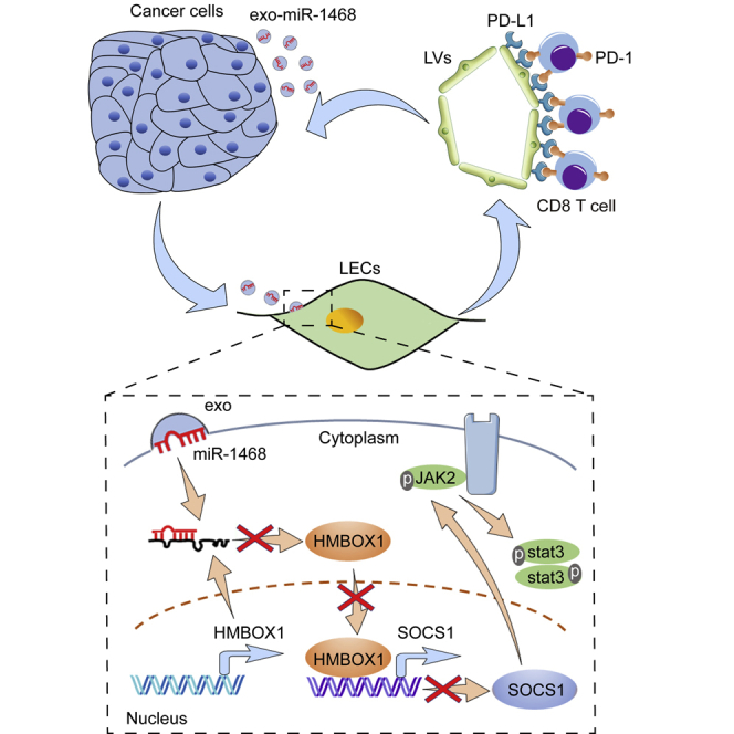
          Graphical abstract

          Abstract

          Our study elucidated a mechanistic model via which cervical cancer-secreted, exosome-encapsulated miR-1468-5p promotes lymphatic PD-L1 upregulation and lymphangiogenesis to impair T cell immunity. Moreover, clinical data revealed that high serum exosomal miR-1468-5p levels correlate with TME immunosuppressive status and poor prognosis in cervical cancer patients.

          Related collections

          Most cited references50

          • Record: found
          • Abstract: found
          • Article: not found

          Global Cancer Statistics 2018: GLOBOCAN Estimates of Incidence and Mortality Worldwide for 36 Cancers in 185 Countries

          This article provides a status report on the global burden of cancer worldwide using the GLOBOCAN 2018 estimates of cancer incidence and mortality produced by the International Agency for Research on Cancer, with a focus on geographic variability across 20 world regions. There will be an estimated 18.1 million new cancer cases (17.0 million excluding nonmelanoma skin cancer) and 9.6 million cancer deaths (9.5 million excluding nonmelanoma skin cancer) in 2018. In both sexes combined, lung cancer is the most commonly diagnosed cancer (11.6% of the total cases) and the leading cause of cancer death (18.4% of the total cancer deaths), closely followed by female breast cancer (11.6%), prostate cancer (7.1%), and colorectal cancer (6.1%) for incidence and colorectal cancer (9.2%), stomach cancer (8.2%), and liver cancer (8.2%) for mortality. Lung cancer is the most frequent cancer and the leading cause of cancer death among males, followed by prostate and colorectal cancer (for incidence) and liver and stomach cancer (for mortality). Among females, breast cancer is the most commonly diagnosed cancer and the leading cause of cancer death, followed by colorectal and lung cancer (for incidence), and vice versa (for mortality); cervical cancer ranks fourth for both incidence and mortality. The most frequently diagnosed cancer and the leading cause of cancer death, however, substantially vary across countries and within each country depending on the degree of economic development and associated social and life style factors. It is noteworthy that high-quality cancer registry data, the basis for planning and implementing evidence-based cancer control programs, are not available in most low- and middle-income countries. The Global Initiative for Cancer Registry Development is an international partnership that supports better estimation, as well as the collection and use of local data, to prioritize and evaluate national cancer control efforts. CA: A Cancer Journal for Clinicians 2018;0:1-31. © 2018 American Cancer Society.
            Bookmark
            • Record: found
            • Abstract: found
            • Article: not found

            Exosome-mediated transfer of mRNAs and microRNAs is a novel mechanism of genetic exchange between cells.

            Exosomes are vesicles of endocytic origin released by many cells. These vesicles can mediate communication between cells, facilitating processes such as antigen presentation. Here, we show that exosomes from a mouse and a human mast cell line (MC/9 and HMC-1, respectively), as well as primary bone marrow-derived mouse mast cells, contain RNA. Microarray assessments revealed the presence of mRNA from approximately 1300 genes, many of which are not present in the cytoplasm of the donor cell. In vitro translation proved that the exosome mRNAs were functional. Quality control RNA analysis of total RNA derived from exosomes also revealed presence of small RNAs, including microRNAs. The RNA from mast cell exosomes is transferable to other mouse and human mast cells. After transfer of mouse exosomal RNA to human mast cells, new mouse proteins were found in the recipient cells, indicating that transferred exosomal mRNA can be translated after entering another cell. In summary, we show that exosomes contain both mRNA and microRNA, which can be delivered to another cell, and can be functional in this new location. We propose that this RNA is called "exosomal shuttle RNA" (esRNA).
              Bookmark
              • Record: found
              • Abstract: found
              • Article: not found

              Predictive correlates of response to the anti-PD-L1 antibody MPDL3280A in cancer patients.

              The development of human cancer is a multistep process characterized by the accumulation of genetic and epigenetic alterations that drive or reflect tumour progression. These changes distinguish cancer cells from their normal counterparts, allowing tumours to be recognized as foreign by the immune system. However, tumours are rarely rejected spontaneously, reflecting their ability to maintain an immunosuppressive microenvironment. Programmed death-ligand 1 (PD-L1; also called B7-H1 or CD274), which is expressed on many cancer and immune cells, plays an important part in blocking the 'cancer immunity cycle' by binding programmed death-1 (PD-1) and B7.1 (CD80), both of which are negative regulators of T-lymphocyte activation. Binding of PD-L1 to its receptors suppresses T-cell migration, proliferation and secretion of cytotoxic mediators, and restricts tumour cell killing. The PD-L1-PD-1 axis protects the host from overactive T-effector cells not only in cancer but also during microbial infections. Blocking PD-L1 should therefore enhance anticancer immunity, but little is known about predictive factors of efficacy. This study was designed to evaluate the safety, activity and biomarkers of PD-L1 inhibition using the engineered humanized antibody MPDL3280A. Here we show that across multiple cancer types, responses (as evaluated by Response Evaluation Criteria in Solid Tumours, version 1.1) were observed in patients with tumours expressing high levels of PD-L1, especially when PD-L1 was expressed by tumour-infiltrating immune cells. Furthermore, responses were associated with T-helper type 1 (TH1) gene expression, CTLA4 expression and the absence of fractalkine (CX3CL1) in baseline tumour specimens. Together, these data suggest that MPDL3280A is most effective in patients in which pre-existing immunity is suppressed by PD-L1, and is re-invigorated on antibody treatment.
                Bookmark

                Author and article information

                Contributors
                Journal
                Mol Ther
                Mol Ther
                Molecular Therapy
                American Society of Gene & Cell Therapy
                1525-0016
                1525-0024
                07 April 2021
                01 January 2021
                : 29
                : 4
                : 1512-1528
                Affiliations
                [1 ]Department of Immunology, School of Basic Medical Sciences, Southern Medical University, Guangdong Provincial Key Laboratory of Proteomics, Guangzhou 510515, China
                [2 ]Department of Obstetrics and Gynecology, The First Affiliated Hospital of Guangzhou Medical University, Guangzhou 510120, China
                [3 ]Department of Gynecology, Guangdong Provincial People’s Hospital, Guangdong Academy of Medical Sciences, Guangzhou 510000, China
                [4 ]Translational and Clinical Research Institute, Faculty of Medical Sciences, Framlington Place, Newcastle University, Newcastle-Upon-Tyne NE2 4HH, UK
                Author notes
                []Corresponding author: Wei Wang, Department of Obstetrics and Gynecology, The First Affiliated Hospital of Guangzhou Medical University, No. 151, Yanjiang Road, Yuexiu District, Guangzhou 510120, China. smugowwang@ 123456126.com
                [∗∗ ]Corresponding author: Sha Wu, Department of Immunology, School of Basic Medical Sciences, Southern Medical University, Guangdong Provincial Key Laboratory of Proteomics, 1838 Guangzhou Avenue North, Baiyun District, Guangzhou 510515, China. shawu99@ 123456outlook.com
                [5]

                These authors contributed equally

                Article
                S1525-0016(20)30727-9
                10.1016/j.ymthe.2020.12.034
                8058488
                33388421
                67f94ca9-6902-4ea8-a293-cd8cfc19d02b
                © 2021 The Authors

                This is an open access article under the CC BY-NC-ND license (http://creativecommons.org/licenses/by-nc-nd/4.0/).

                History
                : 17 August 2020
                : 23 December 2020
                Categories
                Original Article

                Molecular medicine
                Molecular medicine

                Comments

                Comment on this article