11
views
0
recommends
+1 Recommend
0 collections
    0
    shares
      • Record: found
      • Abstract: found
      • Article: found
      Is Open Access

      Efficacy of magnetic resonance imaging for diagnosis of penile fracture: A controlled study

      research-article

      Read this article at

      Bookmark
          There is no author summary for this article yet. Authors can add summaries to their articles on ScienceOpen to make them more accessible to a non-specialist audience.

          Abstract

          Purpose

          To evaluate the diagnostic value of magnetic resonance imaging (MRI) in patients with suspected penile fracture.

          Materials and Methods

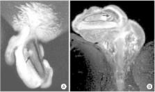
          A total of 122 patients admitted to our inpatient clinic with a suspicion of penile fracture following a recent history of penile trauma and who underwent surgical exploration were included this study. A thorough physical examination, a detailed medical history, description of the trauma, and preoperative International Index of Erectile Function (IIEF) scores were obtained for each patient prior to surgery. Thirty-eight of these patients were evaluated with MRI before the surgical exploration. Intraoperative findings were also recorded. Physical findings and IIEF scores were also recorded at postoperative 6 months.

          Results

          The mean age of our patient group was 36.5±12.3 years. Penile fracture was detected in 105 of 122 patients in whom surgical exploration was performed owing to a suspected diagnosis. The mean time interval from penile trauma to hospital admittance was 9.9±15.1 hours. No cavernosal defect was detected in 9 of 84 patients (10.7%) who were not evaluated with MRI prior to surgery. Compared with surgical exploration, MRI findings showed 100% (30 of 30) sensitivity and 87.5% (7 of 8) specificity in the diagnosis of penile fracture. MRI had a high negative predictive value of 100% (7 of 7) and a positive predictive value of 96.7% (30 of 31) with just 1 misdiagnosed patient.

          Conclusions

          MRI is a reliable diagnostic tool in the diagnosis of penile fractures. Compared to history and physical findings taken all together, the high sensitivity and specificity of this imaging technique can decrease the number of unnecessary surgical explorations.

          Related collections

          Most cited references19

          • Record: found
          • Abstract: found
          • Article: not found

          Fracture of the penis.

          N Eke (2002)
          Sporadic reports of penile fracture give the impression of a rare trauma. The value of diagnostic investigations is doubtful and treatment options are controversial. A Medline search from January 1966 to July 2001 using the terms 'fracture of penis', 'penile trauma' and 'coital injuries' was used to identify full texts of publications on fracture of the penis. Full texts of relevant references from these publications were also identified. Data extracted for review included authors, country and year of publication, number of cases in each report, aetiology, clinical features, investigations, treatment and outcome. In 183 publications 1331 cases were reported between January 1935 and July 2001. Most reports were from the Mediterranean region. The commonest causes were coitus and penile manipulations, especially masturbation. Most patients were in their fourth decade. Clinical features included sudden penile pain, detumescence, voiding difficulties, and penile swelling and deviation. Diagnosis was made mainly on clinical grounds. Associated injuries included urethral rupture. Predisposing factors included excessive force at coitus or manipulation, fibrosclerosis of the tunica albuginea and chronic urethritis. Most authors advocated early surgical repair using absorbable sutures. Complications of the injury included coital difficulty, urethral fistula, penile plaque and erectile dysfunction. Penile fracture is not rare. Radiological investigations are expensive and may delay treatment. Current management favours early surgical exploration to prevent complications.
            Bookmark
            • Record: found
            • Abstract: found
            • Article: not found

            Penile fracture: diagnosis, treatment and outcomes of 150 patients.

            To report the diagnosis, treatment options, and outcomes of 150 patients with suspicion of penile fracture.
              Bookmark
              • Record: found
              • Abstract: found
              • Article: not found

              Sexual function and tunica albuginea wound healing following penile fracture: An 18-year follow-up study of 352 patients from Kermanshah, Iran.

              We present a study on the experiences of penile fracture in an Iranian population. Aim. To determine the long-term outcome of penile fracture. Between April 1990 and May 2008, 373 patients presented with clinical features suggestive of penile fracture. Of these, 11 declined surgery. The remaining 362 were operated upon using a degloving incision. Ten patients had venous injury and 352 had penile fracture. At follow-up visits, in addition to answering our questionnaire, the patients completed the International Index of Erectile Function (IIEF), Erection Hardness Grading Scale (EHGS), and global self-assessment of potency (GSAP). To enhance documentation and to promote transparency, with the patients' permission, their full name and hospital chart number was sent to the journal. Clinical findings and IIEF and EHGS scores. Mean patients' age was 29.6 years. Mean duration of follow-up was 93.6 months. Diagnosis was solely clinical. At presentation, 278 (78.9%) reported no pain. Cavernosography, ultrasonography, or magnetic resonance imaging was not used in any of the patients. Penile fracture was due to taqaandan in 269 patients (76.4%). Patients were treated with surgical exploration and repair within 24 hours of admission, regardless of delay in presentation. A nodule was found at follow-up in 330 patients (93.7%). The painless, mostly proximal nodule was palpated at the floor of the corpora cavernosa, in a deep midline position above the corpus spongiosum. The non-expansive nodule was not associated with erectile dysfunction (ED) or Peyronie's disease. Postoperative complications included mild penile pain in cold weather (two patients), transient wound edema (one patient), mild chordee (four patients), and occasional instability of the erect penis (one patient). Postoperatively, of the 217 patients who had partners, 214 (98.6%) were potent. Mean IIEF ED domain score was 29.8 +/- 1.1. The EHGS score was 4 in 203 and 3 in 11. The GSAP score was 0 in 204, 1 in 8, and 2 in 2. ED in the remaining three could not be explained by penile fracture. Of 10 nonoperated patients, eight (80%) developed ED. Pain is rare in penile fracture. Postoperatively, almost all patients develop a permanent, inconsequential, fibrotic nodule. Our time-tested approach provided excellent long-term sexual function.
                Bookmark

                Author and article information

                Journal
                Investig Clin Urol
                Investig Clin Urol
                ICU
                Investigative and Clinical Urology
                The Korean Urological Association
                2466-0493
                2466-054X
                July 2017
                02 June 2017
                : 58
                : 4
                : 255-260
                Affiliations
                [1 ]Dr. Lütfi Kırdar Education and Research Hospital Urology Clinic, Istanbul, Turkey.
                [2 ]Artvin State Hospital, Artvin, Turkey.
                Author notes
                Corresponding Author: Erkin Saglam. Dr. Lütfi Kırdar Kartal Eğitim ve Araştırma Hastanesi Denizer Cad., Cevizli/Istanbul 34500, Turkey. TEL: +90-216-3068265, FAX: +90-216-3833193, erkinsaglam@ 123456hotmail.com
                Article
                10.4111/icu.2017.58.4.255
                5494349
                28681035
                68591b3b-6de1-4bcb-adbd-c93d14f69076
                © The Korean Urological Association, 2017

                This is an Open Access article distributed under the terms of the Creative Commons Attribution Non-Commercial License ( http://creativecommons.org/licenses/by-nc/4.0) which permits unrestricted non-commercial use, distribution, and reproduction in any medium, provided the original work is properly cited.

                History
                : 22 December 2016
                : 02 March 2017
                Categories
                Original Article
                Sexual Dysfunction/Infertility

                magnetic resonance imaging,penis,rupture
                magnetic resonance imaging, penis, rupture

                Comments

                Comment on this article