Inviting an author to review:
Find an author and click ‘Invite to review selected article’ near their name.
Search for authorsSearch for similar articles
27
views
0
recommends
+1 Recommend
0 collections
    0
    shares
      • Record: found
      • Abstract: found
      • Article: found
      Is Open Access

      Effects of Omega-3 Fatty Acids on Erectile Dysfunction in a Rat Model of Atherosclerosis-induced Chronic Pelvic Ischemia

      research-article

      Read this article at
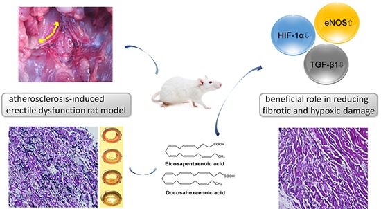
      Bookmark
          There is no author summary for this article yet. Authors can add summaries to their articles on ScienceOpen to make them more accessible to a non-specialist audience.

          Abstract

          The aim of this study was to investigate whether the omega-3 fatty acids help to improve erectile function in an atherosclerosis-induced erectile dysfunction rat model. A total of 20 male Sprague-Dawley rats at age 8 weeks were divided into three groups: Control group (n = 6, untreated sham operated rats), Pathologic group (n = 7, untreated rats with chronic pelvic ischemia [CPI]), and Treatment group (n = 7, CPI rats treated with omega-3 fatty acids). For the in vivo study, electrical stimulation of the cavernosal nerve was performed and erectile function was measured in all groups. Immunohistochemical antibody staining was performed for transforming growth factor beta-1 (TGF-β1), endothelial nitric oxide synthase (eNOS), and hypoxia inducible factor 1-alpha (HIF-1α). In vivo measurement of erectile function in the Pathologic group showed significantly lower values than those in the Control group, whereas the Treatment group showed significantly improved values in comparison with those in the Pathologic group. The results of western blot analysis revealed that systemically administered omega-3 fatty acids ameliorated the cavernosal molecular environment. Our study suggests that omega-3 fatty acids improve intracavernosal pressure and have a beneficial role against pathophysiological consequences such as fibrosis or hypoxic damage on a CPI rat model, which represents a structural erectile dysfunction model.

          Graphical Abstract

          Related collections

          Most cited references21

          • Record: found
          • Abstract: found
          • Article: not found

          Long chain omega-3 fatty acids and cardiovascular disease: a systematic review.

          Cardiovascular disease remains the commonest health problem in developed countries, and residual risk after implementing all current therapies is still high. The use of marine omega-3 fatty acids (DHA and EPA) has been recommended to reduce cardiovascular risk by multiple mechanisms. To update the current evidence on the influence of omega-3 on the rate of cardiovascular events. We used the MEDLINE and EMBASE databases to identify clinical trials and randomized controlled trials of omega-3 fatty acids (with quantified quantities) either in capsules or in dietary intake, compared to placebo or usual diet, equal to or longer than 6 months, and written in English. The primary outcome was a cardiovascular event of any kind and secondary outcomes were all-cause mortality, cardiac death and coronary events. We used RevMan 5·1 (Mantel-Haenszel method). Heterogeneity was assessed by the I2 and Chi2 tests. We included 21 of the 452 pre-selected studies. We found an overall decrease of risk of suffering a cardiovascular event of any kind of 10 % (OR 0·90; [0·85-0·96], p = 0·001), a 9 % decrease of risk of cardiac death (OR 0·91; [0·83-0·99]; p = 0·03), a decrease of coronary events (fatal and non-fatal) of 18 % (OR 0·82; [0·75-0·90]; p < 1 × 10⁻⁴), and a trend to lower total mortality (5 % reduction of risk; OR 0·95; [0·89-1·02]; p = 0·15. Most of the studies analyzed included persons with high cardiovascular risk. marine omega-3 fatty acids are effective in preventing cardiovascular events, cardiac death and coronary events, especially in persons with high cardiovascular risk.
            Bookmark
            • Record: found
            • Abstract: found
            • Article: not found

            Histological alterations in cavernous tissue after radical prostatectomy.

            Radical prostatectomy often results in erectile dysfunction because of lesions to the erectile nerves. In this study we evaluated histomorphological alterations in cavernous smooth muscle and collagen content after radical prostatectomy. A total of 19 patients between 57 and 69 years old with prostate adenocarcinoma and normal erectile function, as reported and validated by RigiScan (UroHealth Systems, Laguna Niguel, California) testing, underwent corpora cavernosa biopsy in the operating room before radical prostatectomy, and 2 and 12 months after surgery. No patient underwent hormone therapy before or after surgery and none was diabetic. Elastic fibers (manual counting), muscle specific actin (immunostaining) and collagen content (computerized morphometric imaging) were measured in the 3 biopsies. In all cases the first postoperative histological assessment revealed some disorganization. Trabecular elastic fibers (p <0.0003) and smooth muscle fibers were decreased and collagen content was significantly increased (p <0.0003) compared with preoperative biopsies. One year after surgery elastic fibers (p <0.0003) and smooth muscle fibers were decreased and collagen content was significantly increased (p <0.0003) compared with the first postoperative biopsy. Moreover, organized collagen and trabecular protocollagen deposits were increased. Progressive fibrosis in the corpora cavernosa after radical prostatectomy probably results from denervation and/or an ischemic process, which is caused in turn by the ligation of anomalous pudendal artery branches or of venous plexuses that drain to or from the corpora cavernosa. Fibrosis and the subsequent loss in elasticity and function of erectile tissue probably together cause erectile dysfunction.
              Bookmark
              • Record: found
              • Abstract: found
              • Article: not found

              The omega-3 fatty acid docosahexaenoate reduces cytokine-induced expression of proatherogenic and proinflammatory proteins in human endothelial cells.

              The mechanisms by which dietary fatty acids can modulate atherogenesis and inflammation are poorly understood. Induction in endothelial cells of adhesion molecules for circulating leukocytes and of inflammatory mediators by cytokines probably contributes to the early phases of atherogenesis and inflammation. We report here that incorporation into cellular lipids of docosahexaenoic acid (DHA), a specific fatty acid of the omega 3 family, decreases cytokine-induced expression of endothelial leukocyte adhesion molecules, secretion of inflammatory mediators, and leukocyte adhesion to cultured endothelial cells. DHA, but not eicosapentaenoic acid, decreased in a dose- and time-dependent fashion the expression of vascular cell adhesion molecule 1 (VCAM-1) induced by interleukin (IL)-1, tumor necrosis factor (TNF), IL-4, or bacterial lipopolysaccharide, with half-maximum inhibition at < 10 mumol/L. This reduction required prolonged (24- to 96-hour) exposure of endothelial cells to DHA and correlated with the degree of DHA incorporation into cellular lipids. DHA also limited cytokine-stimulated endothelial cell expression of E-selectin and intercellular adhesion molecule 1 and the secretion of IL-6 and IL-8 into the medium but not the surface expression of constitutive surface molecules. Cyclooxygenase inhibition did not block the effect of DHA on VCAM-1. In parallel with reduced surface VCAM-1 protein expression, DHA reduced VCAM-1 mRNA induction by IL-1 or TNF. DHA treatment also reduced the adhesion of human monocytes and of monocytic U937 cells to cytokine-stimulated endothelial cells. These properties of DHA may contribute to antiatherogenic and anti-inflammatory effects of omega 3 fatty acids.
                Bookmark

                Author and article information

                Journal
                J Korean Med Sci
                J. Korean Med. Sci
                JKMS
                Journal of Korean Medical Science
                The Korean Academy of Medical Sciences
                1011-8934
                1598-6357
                April 2016
                22 February 2016
                : 31
                : 4
                : 585-589
                Affiliations
                Department of Urology, Korea University College of Medicine, Seoul, Korea.
                Author notes
                Address for Correspondence: Du Geon Moon, MD. Department of Urology, Korea University Guro Hospital, Korea University College of Medicine, 148 Gurodong-ro, Guro-gu, Seoul 08308, Korea. dgmoon@ 123456korea.ac.kr
                Author information
                http://orcid.org/0000-0002-6745-1776
                http://orcid.org/0000-0002-9031-9845
                Article
                10.3346/jkms.2016.31.4.585
                4810342
                27051243
                6898172c-c3fc-4a36-b1b9-43483c61948d
                © 2016 The Korean Academy of Medical Sciences.

                This is an Open Access article distributed under the terms of the Creative Commons Attribution Non-Commercial License ( http://creativecommons.org/licenses/by-nc/4.0/) which permits unrestricted non-commercial use, distribution, and reproduction in any medium, provided the original work is properly cited.

                History
                : 13 September 2015
                : 23 December 2015
                Categories
                Original Article
                Urology

                Medicine
                erectile dysfunction,fatty acids, omega-3,ischemia,rats
                Medicine
                erectile dysfunction, fatty acids, omega-3, ischemia, rats

                Comments

                Comment on this article