48
views
0
recommends
+1 Recommend
0 collections
    0
    shares
      • Record: found
      • Abstract: found
      • Article: found
      Is Open Access

      Global and Ocular Hypothermic Preconditioning Protect the Rat Retina from Ischemic Damage

      research-article

      Read this article at

      Bookmark
          There is no author summary for this article yet. Authors can add summaries to their articles on ScienceOpen to make them more accessible to a non-specialist audience.

          Abstract

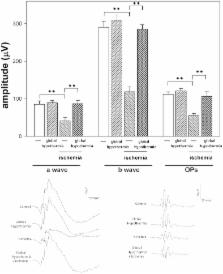
          Retinal ischemia could provoke blindness. At present, there is no effective treatment against retinal ischemic damage . Strong evidence supports that glutamate is implicated in retinal ischemic damage. We investigated whether a brief period of global or ocular hypothermia applied 24 h before ischemia (i.e. hypothermic preconditioning, HPC) protects the retina from ischemia/reperfusion damage, and the involvement of glutamate in the retinal protection induced by HPC. For this purpose, ischemia was induced by increasing intraocular pressure to 120 mm Hg for 40 min. One day before ischemia, animals were submitted to global or ocular hypothermia (33°C and 32°C for 20 min, respectively) and fourteen days after ischemia, animals were subjected to electroretinography and histological analysis. Global or ocular HPC afforded significant functional (electroretinographic) protection in eyes exposed to ischemia/reperfusion injury. A marked alteration of the retinal structure and a decrease in retinal ganglion cell number were observed in ischemic retinas, whereas global or ocular HPC significantly preserved retinal structure and ganglion cell count. Three days after ischemia, a significant decrease in retinal glutamate uptake and glutamine synthetase activity was observed, whereas ocular HPC prevented the effect of ischemia on these parameters. The intravitreal injection of supraphysiological levels of glutamate induced alterations in retinal function and histology which were significantly prevented by ocular HPC. These results support that global or ocular HPC significantly protected retinal function and histology from ischemia/reperfusion injury, probably through a glutamate-dependent mechanism.

          Related collections

          Most cited references47

          • Record: found
          • Abstract: found
          • Article: not found

          Retinal ischemia: mechanisms of damage and potential therapeutic strategies.

          Retinal ischemia is a common cause of visual impairment and blindness. At the cellular level, ischemic retinal injury consists of a self-reinforcing destructive cascade involving neuronal depolarisation, calcium influx and oxidative stress initiated by energy failure and increased glutamatergic stimulation. There is a cell-specific sensitivity to ischemic injury which may reflect variability in the balance of excitatory and inhibitory neurotransmitter receptors on a given cell. A number of animal models and analytical techniques have been used to study retinal ischemia, and an increasing number of treatments have been shown to interrupt the "ischemic cascade" and attenuate the detrimental effects of retinal ischemia. Thus far, however, success in the laboratory has not been translated to the clinic. Difficulties with the route of administration, dosage, and adverse effects may render certain experimental treatments clinically unusable. Furthermore, neuroprotection-based treatment strategies for stroke have so far been disappointing. However, compared to the brain, the retina exhibits a remarkable natural resistance to ischemic injury, which may reflect its peculiar metabolism and unique environment. Given the increasing understanding of the events involved in ischemic neuronal injury it is hoped that clinically effective treatments for retinal ischemia will soon be available.
            Bookmark
            • Record: found
            • Abstract: found
            • Article: not found

            Small differences in intraischemic brain temperature critically determine the extent of ischemic neuronal injury.

            We have tested whether small intraischemic variations in brain temperature influence the outcome of transient ischemia. To measure brain temperature, a thermocouple probe was placed stereotaxically into the left dorsolateral striatum of rats prior to 20 min of four-vessel occlusion. Rectal temperature was maintained at 36-37 degrees C by a heating lamp, and striatal temperature prior to ischemia was 36 degrees C in all animals. Six animal subgroups were investigated, including rats whose intraischemic striatal brain temperature was not regulated, or was maintained at 33, 34, 36, or 39 degrees C. Postischemic brain temperature was regulated at 36 degrees C, except for one group in which brain temperature was lowered from 36 degrees C to 33 degrees C during the first hour of recirculation. Energy metabolites were measured at the end of the ischemic insult, and histopathological evaluation was carried out at 3 days after ischemia. Intraischemic variations in brain temperature had no significant influence on energy metabolite levels measured at the conclusion of ischemia: Severe depletion of brain ATP, phosphocreatine, glucose, and glycogen and elevation of lactate were observed to a similar degree in all experimental groups. The histopathological consequences of ischemia, however, were markedly influenced by variations in intraischemic brain temperature. In the hippocampus, CA1 neurons were consistently damaged at 36 degrees C, but not at 34 degrees C. Within the dorsolateral striatum, ischemic cell change was present in 100% of the hemispheres at 36 degrees C, but in only 50% at 34 degrees C. Ischemic neurons within the central zone of striatum were not observed in any rats at 34 degrees C, but in all rats at 36 degrees C. In rats whose striatal temperature was not controlled, brain temperature fell from 36 to 30-31 degrees C during the ischemic insult. In this group, no ischemic cell change was seen within striatal areas and was only inconsistently documented within the CA1 hippocampal region. These results demonstrate that (a) rectal temperature unreliably reflects brain temperature during ischemia; (b) despite severe depletion of brain energy metabolites during ischemia at all temperatures, small increments of intraischemic brain temperature markedly accentuate histopathological changes following 3-day survival; and (c) brain temperature must be controlled above 33 degrees C in order to ensure a consistent histopathological outcome. Lowering of the brain temperature by only a few degrees during ischemia confers a marked protective effect.
              Bookmark
              • Record: found
              • Abstract: found
              • Article: not found

              Treatment of traumatic brain injury with moderate hypothermia.

              Traumatic brain injury initiates several metabolic processes that can exacerbate the injury. There is evidence that hypothermia may limit some of these deleterious metabolic responses. In a randomized, controlled trial, we compared the effects of moderate hypothermia and normothermia in 82 patients with severe closed head injuries (a score of 3 to 7 on the Glasgow Coma Scale). The patients assigned to hypothermia were cooled to 33 degrees C a mean of 10 hours after injury, kept at 32 degrees to 33 degrees C for 24 hours, and then rewarmed. A specialist in physical medicine and rehabilitation who was unaware of the treatment assignments evaluated the patients 3, 6, and 12 months later with the use of the Glasgow Outcome Scale. The demographic characteristics and causes and severity of injury were similar in the hypothermia and normothermia groups. At 12 months, 62 percent of the patients in the hypothermia group and 38 percent of those in the normothermia group had good outcomes (moderate, mild, or no disabilities). The adjusted risk ratio for a bad outcome in the hypothermia group was 0.5 (95 percent confidence interval, 0.2 to 1.2). Hypothermia did not improve the outcomes in the patients with coma scores of 3 or 4 on admission. Among the patients with scores of 5 to 7, hypothermia was associated with significantly improved outcomes at 3 and 6 months (adjusted risk ratio for a bad outcome, 0.2; 95 percent confidence interval, 0.1 to 0.9 at both intervals), although not at 12 months (risk ratio, 0.3; 95 percent confidence interval, 0.1 to 1.0). Treatment with moderate hypothermia for 24 hours in patients with severe traumatic brain injury and coma scores of 5 to 7 on admission hastened neurologic recovery and may have improved the outcome.
                Bookmark

                Author and article information

                Contributors
                Role: Editor
                Journal
                PLoS One
                PLoS ONE
                plos
                plosone
                PLoS ONE
                Public Library of Science (San Francisco, USA )
                1932-6203
                2013
                23 April 2013
                : 8
                : 4
                : e61656
                Affiliations
                [1]Laboratory of Retinal Neurochemistry and Experimental Ophthalmology, Department of Human Biochemistry, School of Medicine, University of Buenos Aires/CEFyBO, CONICET, Buenos Aires, Argentina
                Eye Hospital, Charité, Germany
                Author notes

                Competing Interests: The authors have declared that no competing interests exist.

                Conceived and designed the experiments: EMS DD RER. Performed the experiments: EMS DD MB MFGF MC. Analyzed the data: EMS DD RER. Wrote the paper: EMS RER.

                Article
                PONE-D-12-38429
                10.1371/journal.pone.0061656
                3633982
                23626711
                68a9ac3e-70ae-4608-a05d-83fbbd822f78
                Copyright @ 2013

                This is an open-access article distributed under the terms of the Creative Commons Attribution License, which permits unrestricted use, distribution, and reproduction in any medium, provided the original author and source are credited.

                History
                : 4 December 2012
                : 12 March 2013
                Page count
                Pages: 13
                Funding
                This research was supported by grants from the Agencia Nacional de Promoción Científica y Tecnológica; The University of Buenos Aires; and Consejo Nacional de Investigaciones Científicas y Técnicas, Argentina. The funders had no role in study design, data collection and analysis, decision to publish, or preparation of the manuscript.
                Categories
                Research Article
                Biology
                Anatomy and Physiology
                Ocular System
                Biochemistry
                Neurochemistry
                Neuroscience
                Sensory Systems
                Visual System
                Cellular Neuroscience
                Neurobiology of Disease and Regeneration
                Medicine
                Anatomy and Physiology
                Ocular System
                Ophthalmology
                Retinal Disorders

                Uncategorized
                Uncategorized

                Comments

                Comment on this article