162
views
0
recommends
+1 Recommend
0 collections
    0
    shares
      • Record: found
      • Abstract: found
      • Article: found
      Is Open Access

      A Case Presentation: Decidualized Endometrioma Mimicking Ovarian Cancer during Pregnancy

      case-report

      Read this article at

      Bookmark
          There is no author summary for this article yet. Authors can add summaries to their articles on ScienceOpen to make them more accessible to a non-specialist audience.

          Abstract

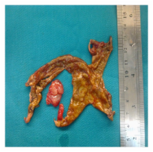
          During pregnancy, masses that are larger than 5 cm and appearing in the Doppler ultrasonography as having increased blood flow, echoes of heterogeneous density, and containing solid components are suspicious for malignancy; however, differential diagnosis of decidualized endometriomas should also be considered. The patient was an 8 weeks pregnant primigravida. The ultrasonographic evaluation showed a cystic mass of size 65 × 57 mm in the left ovary that was well circumscribed, heterogeneous, with highly dense internal echo, and containing a solid component of size 8 × 14 mm. In the 12th week, the ultrasonographic examination revealed an increase in the size of the mass and increased arterial blood flow in the mass. The patient underwent surgery. It was observed that both ovaries were adherent in the Douglas pouch and that the left ovary contained an endometrioma of size 8cm. While the capsule was being peeled, lesions of soft density, with irregular surfaces, and with adhesion in the Douglas pouch were observed. The results of the frozen section revealed decidualized endometrioma and decidual structures. Even in pregnant women when adnexal masses are encountered and the ultrasonography, Doppler, MRI, and CA 125 level analysis still do not favor endometriosis, decidualized endometrioma should be considered in the differential diagnosis.

          Related collections

          Most cited references20

          • Record: found
          • Abstract: found
          • Article: not found

          Malignant neoplasms arising in endometriosis.

          Ten cases of malignant tumors arising in foci of gonadal and extragonadal endometriosis are reported and added to 195 previously reported cases from the English literature. The ovary was the primary site in 165 (78.7%) of the cases, whereas extragonadal sites represented 44 (21.3%). Endometrioid adenocarcinomas accounted for 69% of the lesions, clear-cell carcinomas 13.5%, sarcomas 11.6%, and rare cell types 6%. Extragonadal lesions were mostly endometrioid tumors (66%) and sarcomas (25%). Tumors arising in endometriosis were predominantly low grade and confined to the site of origin. Radiation therapy was often able to control completely tumors limited to the pelvis, but was not beneficial in metastatic disease. Only one patient had a response to chemotherapy. Fourteen patients received postoperative progestin therapy, with a 77% 5-year survival. Follow-up has been reported in 86 patients. The tumor was either confined to the ovary (57), confined to the extragonadal site of origin (11), or spread throughout the peritoneal cavity (18). With each of these situations, the 5-year survival was 65, 100, and 10%, respectively. Fourteen patients had malignant transformation in endometriosis associated with presumed estrogenic stimulation; most lesions (69%) were well differentiated and the 5-year survival was 82%. After surgical resection, we recommend that progestin therapy be included in the treatment of cancer arising in endometriosis. The actual frequency of malignancy arising in endometriosis may be higher than reported.
            Bookmark
            • Record: found
            • Abstract: found
            • Article: not found

            Validation study of nonsurgical diagnosis of endometriosis.

            To determine whether the surgical diagnosis of endometriosis can be predicted using symptoms, signs, and ultrasound findings. Prospective study (study sample); retrospective record review (test sample). Hospital of Desio (study sample) and Mangiagalli Hospital (test sample), Italy. Ninety women scheduled to undergo laparoscopy or laparotomy (study sample); 120 women who underwent laparoscopy (test sample). The study sample group was interviewed before surgery about infertility and dysmenorrhea, dyspareunia, and noncyclic pelvic pain and each member had a pelvic examination and a transvaginal ultrasound. At surgery, endometriosis was noted. For the test sample, the same information was abstracted from medical records after laparoscopy. The ability of symptoms, signs, and ultrasound to predict endometriosis at surgery. A classification tree was developed with the study sample and evaluated with the test sample. Ovarian endometriosis, but not nonovarian endometriosis, could be reliably predicted with noninvasive tools. Ultrasound and examination best predicted ovarian endometriosis, correctly classifying 100% of cases with no false positive diagnoses in the study sample. Similar results were found in the test sample. Noninvasive tools may be used to identify women with ovarian, but not nonovarian endometriosis, with excellent agreement with surgical diagnosis.
              Bookmark
              • Record: found
              • Abstract: not found
              • Article: not found

              Malignant tumors arising in endometriosis.

                Bookmark

                Author and article information

                Journal
                Case Rep Obstet Gynecol
                Case Rep Obstet Gynecol
                CRIM.OBGYN
                Case Reports in Obstetrics and Gynecology
                Hindawi Publishing Corporation
                2090-6684
                2090-6692
                2013
                14 April 2013
                : 2013
                : 728291
                Affiliations
                Department of Obstetrics and Gynecology, Faculty of Medicine, Selcuk University, Selcuklu, Konya 42250, Turkey
                Author notes

                Academic Editors: B. Reime and R. Shaco-Levy

                Author information
                https://orcid.org/0000-0003-2208-8712
                Article
                10.1155/2013/728291
                3639707
                23662226
                6919fb3e-f3e8-4450-9376-a5fb00deed8a
                Copyright © 2013 Aybike Tazegül et al.

                This is an open access article distributed under the Creative Commons Attribution License, which permits unrestricted use, distribution, and reproduction in any medium, provided the original work is properly cited.

                History
                : 5 February 2013
                : 7 March 2013
                Categories
                Case Report

                Obstetrics & Gynecology
                Obstetrics & Gynecology

                Comments

                Comment on this article