0
views
0
recommends
+1 Recommend
0 collections
    0
    shares
      • Record: found
      • Abstract: found
      • Article: found
      Is Open Access

      HASTE MRI sequence findings correlate with loss of deep pain perception in dogs with thoracolumbar disc extrusion

      research-article
      1 , , 1
      Veterinary Medicine and Science
      John Wiley and Sons Inc.
      intervertebral disc extrusion, MRI, spine

      Read this article at

      Bookmark
          There is no author summary for this article yet. Authors can add summaries to their articles on ScienceOpen to make them more accessible to a non-specialist audience.

          Abstract

          Background

          Thoracolumbar intervertebral disc extrusion (TL IVDE) is a common reason for the veterinary hospital admission. Various imaging factors including degree and length of compression have been tested for correlation with clinical severity, but no reliable correlation has been found. Half‐Fourier acquisition single‐shot turbo spin echo (HASTE) magnetic resonance imaging (MRI) sequences highlight the dorsal and ventral cerebrospinal fluid (CSF) columns and have been used to demonstrate spinal cord swelling in dogs with TL IVDE. This has been used as a predictor of progressive ascending‐descending myelomalacia but has not been correlated with neurological grade.

          Objective

          This study aims to investigate the correlation between the attenuation of CSF HASTE signal and clinical severity in dogs suffering from TL disc extrusions.

          Methods

          Dogs less than 15 kg who were non‐ambulatory due to suspected TL IVDE were prospectively recruited for a study into conservative management. MRI studies were undertaken under sedation including HASTE sequences. The ratio of the length of CSF attenuation to the length of the L2 vertebra was calculated and correlated with clinical severity.

          Results

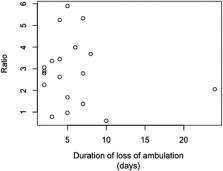
          Twenty dogs met the inclusion criteria. No statistically significant difference was demonstrated between the mean CSF attenuation and neurological grade ( p = 0.17 but there was a significant difference in the mean CSF attenuation in those who retained deep pain perception and those who did not ( p = 0.02). Time to loss of ambulation was also found to not be correlated with CSF attenuation ( p = 0.95).

          Conclusion

          In conclusion, the length of CSF attenuation of HASTE MRI sequences in dogs less than 15 kg suffering from IVDEs may be correlated with a loss of deep pain perception.

          Abstract

          Dogs who were non‐ambulatory due to suspected thoracolumbar intervertebral disc extrusion underwent MRI studies including half‐Fourier acquisition single‐shot turbo spin echo sequences. The ratio of the length of CSF attenuation to the length of the L2 vertebra was calculated and correlated with clinical severity. This showed that the length of CSF attenuation sequences may be correlated with a loss of deep pain perception.

          Related collections

          Most cited references28

          • Record: found
          • Abstract: found
          • Article: not found

          Intervertebral disc disease in dogs.

          Intervertebral disc herniation is a common cause of neurologic dysfunction in dogs. This article reviews the anatomy, pathophysiology, diagnostic imaging, treatment options, and prognosis for canine cervical and thoracolumbar intervertebral disc disease. The extensive literature pertinent to intervertebral disc disease is reviewed with the goal of summarizing the information available to help clinicians in their decision making. Copyright 2010 Elsevier Inc. All rights reserved.
            Bookmark
            • Record: found
            • Abstract: found
            • Article: not found

            Prognostic value of magnetic resonance imaging in dogs with paraplegia caused by thoracolumbar intervertebral disk extrusion: 77 cases (2000-2003).

            To determine whether magnetic resonance imaging findings in dogs with paraplegia caused by thoracolumbar intervertebral disk extrusion were predictive of clinical outcome. Retrospective case series. 77 dogs. Medical records and magnetic resonance images were reviewed; clinical outcome was classified as successful (regained ability to walk with no more than mild neurologic deficits) or unsuccessful (severe neurologic deficits persisted). The prognostic value of magnetic resonance imaging was compared with prognostic value of deep pain perception, duration of clinical signs, and rate of onset of clinical signs. 33 (43%) dogs had areas of hyperintensity of the spinal cord greater than or equal to the length of the L2 vertebral body on T2-weighted magnetic resonance images. All 44 dogs without areas of hyperintensity on T2-weighted images had a successful outcome, but only 18 of the 33 (55%) dogs with an area of hyperintensity did. Only 5 of 16 dogs with an area of hyperintensity that had also lost deep pain perception had a successful outcome. The odds ratio for an unsuccessful outcome for a dog with an area of hyperintensity (29.87) was higher than the odds ratio for a dog that had lost deep pain perception (5.24). Duration and rate of onset of clinical signs were not associated with clinical outcome. Findings suggest that results of magnetic resonance imaging can be used to predict clinical outcome in dogs with paraplegia caused by intervertebral disk extrusion.
              Bookmark
              • Record: found
              • Abstract: found
              • Article: not found

              Factors associated with recovery from paraplegia in dogs with loss of pain perception in the pelvic limbs following intervertebral disk herniation.

              To investigate associations between recovery of locomotion and putative prognostic factors in dogs with loss of deep pain perception in the pelvic limbs caused by intervertebral disk herniation (IVDH).
                Bookmark

                Author and article information

                Contributors
                shk47@cam.ac.uk
                Journal
                Vet Med Sci
                Vet Med Sci
                10.1002/(ISSN)2053-1095
                VMS3
                Veterinary Medicine and Science
                John Wiley and Sons Inc. (Hoboken )
                2053-1095
                27 October 2022
                March 2023
                : 9
                : 2 ( doiID: 10.1002/vms3.v9.2 )
                : 604-608
                Affiliations
                [ 1 ] Queen's Veterinary School Hospital Cambridge UK
                Author notes
                [*] [* ] Correspondence

                Sam Khan, Queen's Veterinary School Hospital, Madingley Road, Cambridge, CB3 0ES, UK.

                Email: shk47@ 123456cam.ac.uk

                Author information
                https://orcid.org/0000-0002-4254-858X
                https://orcid.org/0000-0001-6326-5573
                Article
                VMS3974
                10.1002/vms3.974
                10029906
                36303241
                6ab70dfe-a769-4715-8c6d-b3d3b3c1029f
                © 2022 The Authors. Veterinary Medicine and Science published by John Wiley & Sons Ltd.

                This is an open access article under the terms of the http://creativecommons.org/licenses/by-nc-nd/4.0/ License, which permits use and distribution in any medium, provided the original work is properly cited, the use is non‐commercial and no modifications or adaptations are made.

                History
                Page count
                Figures: 5, Tables: 0, Pages: 5, Words: 3768
                Funding
                Funded by: Petsavers and the Kennel Club Charitable Trust
                Categories
                Original Article
                DOGS
                Original Articles
                Custom metadata
                2.0
                March 2023
                Converter:WILEY_ML3GV2_TO_JATSPMC version:6.2.6 mode:remove_FC converted:21.03.2023

                intervertebral disc extrusion,mri,spine
                intervertebral disc extrusion, mri, spine

                Comments

                Comment on this article