1
views
0
recommends
+1 Recommend
0 collections
    0
    shares
      • Record: found
      • Abstract: found
      • Article: found
      Is Open Access

      Fetal Pain in the First Trimester

      research-article
      , MD, MS 1 ,
      The Linacre Quarterly
      SAGE Publications
      fetal pain, fetal analgesia, fetal anesthesia, fetal nociception, fetal awareness

      Read this article at

      Bookmark
          There is no author summary for this article yet. Authors can add summaries to their articles on ScienceOpen to make them more accessible to a non-specialist audience.

          Abstract

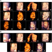
          Fetal pain perception has important implications for fetal surgery, as well as for abortion. Current neuroscientific evidence indicates the possibility of fetal pain perception during the first trimester (<14 weeks gestation). Evidence for this conclusion is based on the following findings: (1) the neural pathways for pain perception via the cortical subplate are present as early as 12 weeks gestation, and via the thalamus as early as 7–8 weeks gestation; (2) the cortex is not necessary for pain to be experienced; (3) consciousness is mediated by subcortical structures, such as the thalamus and brainstem, which begin to develop during the first trimester; (4) the neurochemicals in utero do not cause fetal unconsciousness; and (5) the use of fetal analgesia suppresses the hormonal, physiologic, and behavioral responses to pain, avoiding the potential for both short- and long-term sequelae. As the medical evidence has shifted in acknowledging fetal pain perception prior to viability, there has been a gradual change in the fetal pain debate, from disputing the existence of fetal pain to debating the significance of fetal pain. The presence of fetal pain creates tension in the practice of medicine with respect to beneficence and nonmaleficence.

          Related collections

          Most cited references127

          • Record: found
          • Abstract: found
          • Article: not found

          The revised International Association for the Study of Pain definition of pain: concepts, challenges, and compromises

          The current International Association for the Study of Pain (IASP) definition of pain as "An unpleasant sensory and emotional experience associated with actual or potential tissue damage, or described in terms of such damage" was recommended by the Subcommittee on Taxonomy and adopted by the IASP Council in 1979. This definition has become accepted widely by health care professionals and researchers in the pain field and adopted by several professional, governmental, and nongovernmental organizations, including the World Health Organization. In recent years, some in the field have reasoned that advances in our understanding of pain warrant a reevaluation of the definition and have proposed modifications. Therefore, in 2018, the IASP formed a 14-member, multinational Presidential Task Force comprising individuals with broad expertise in clinical and basic science related to pain, to evaluate the current definition and accompanying note and recommend whether they should be retained or changed. This review provides a synopsis of the critical concepts, the analysis of comments from the IASP membership and public, and the committee's final recommendations for revisions to the definition and notes, which were discussed over a 2-year period. The task force ultimately recommended that the definition of pain be revised to "An unpleasant sensory and emotional experience associated with, or resembling that associated with, actual or potential tissue damage," and that the accompanying notes be updated to a bulleted list that included the etymology. The revised definition and notes were unanimously accepted by the IASP Council early this year.
            Bookmark
            • Record: found
            • Abstract: found
            • Article: not found

            Development of the human cerebral cortex: Boulder Committee revisited.

            In 1970 the Boulder Committee described the basic principles of the development of the CNS, derived from observations on the human embryonic cerebrum. Since then, numerous studies have significantly advanced our knowledge of the timing, sequence and complexity of developmental events, and revealed important inter-species differences. We review current data on the development of the human cerebral cortex and update the classical model of how the structure that makes us human is formed.
              Bookmark
              • Record: found
              • Abstract: found
              • Article: not found

              Developmental history of the transient subplate zone in the visual and somatosensory cortex of the macaque monkey and human brain.

              The cytological organization and the timetable of emergence and dissolution of the transient subplate zone subjacent to the developing visual and somatosensory cortex were studied in a series of human and monkey fetal brains. Cerebral walls processed with Nissl, Golgi, electron-microscopic, and histochemical methods show that this zone consists of migratory and postmigratory neurons, growth cones, loosely arranged axons, dendrites, synapses, and glial cells. In both species the subplate zone becomes visible at the beginning of the mid-third of gestation as a cell-poor/fiber-rich layer situated between the intermediate zone and the developing cortical plate. The subplate zone appears earlier in the somatosensory than in the visual area and reaches maximal width at the beginning of the last third of gestation in both regions. At the peak of its size the ratio between the width of the subplate zone and cortical plate in the somatosensory cortex is 2:1 in monkey and 4:1 in man while in the occipital lobe these structures have about equal width in both species. The dissolution of the subplate zone begins during the last third of gestation with degeneration of some subplate neurons and the relocation of fiber terminals into the cortex. The subplate zone disappears faster in the visual than in the somatosensory area. The present results together with our previous findings support the hypothesis that the subplate zone may serve as a "waiting" compartment for transient cellular interactions and a substrate for competition, segregation, and growth of afferents originated sequentially from the brain stem, basal forebrain, thalamus, and from the ipsi- and contralateral cerebral hemisphere. After a variable and partially overlapping time period, these fibers enter the cortical plate while the subplate zone disappears leaving only a vestige of cells scattered throughout the subcortical white matter. A comparison between species indicates that the size and duration of the subplate zone increases during mammalian evolution and culminates in human fetuses concomitantly with an enlargement of cortico-cortical fiber systems. The regional difference in the size, pattern, and resolution of the subplate zone correlates also with the pattern of cerebral convolutions. Our findings indicate that, contrary to prevailing notions, the subplate may not be a vestige of the phylogenetically old network but a transient embryonic structure that expanded during evolution to subserve the increasing number of its connections.
                Bookmark

                Author and article information

                Journal
                Linacre Q
                Linacre Q
                splqr
                LQR
                The Linacre Quarterly
                SAGE Publications (Sage CA: Los Angeles, CA )
                0024-3639
                2050-8549
                6 December 2021
                February 2022
                6 December 2021
                : 89
                : 1
                : 73-100
                Affiliations
                [1 ]University of Mary, Bismarck, ND, USA
                Author notes
                [*]Bridget Thill, MD, MS, University of Mary, 7500 University Dr, Bismarck, ND 58504, USA. Email: bathill1@ 123456umary.edu
                Author information
                https://orcid.org/0000-0002-7363-4560
                Article
                10.1177_00243639211059245
                10.1177/00243639211059245
                8935428
                35321491
                6ae6e70d-67ac-43ec-9e86-c604ed5eed87
                © Catholic Medical Association 2021

                This article is distributed under the terms of the Creative Commons Attribution-NonCommercial 4.0 License ( https://creativecommons.org/licenses/by-nc/4.0/) which permits non-commercial use, reproduction and distribution of the work without further permission provided the original work is attributed as specified on the SAGE and Open Access page ( https://us.sagepub.com/en-us/nam/open-access-at-sage).

                History
                Categories
                Original Research
                Custom metadata
                ts10

                fetal pain,fetal analgesia,fetal anesthesia,fetal nociception,fetal awareness

                Comments

                Comment on this article