26
views
0
recommends
+1 Recommend
0 collections
    0
    shares
      • Record: found
      • Abstract: found
      • Article: found
      Is Open Access

      Diversity in Protein Profiles of Individual Calcium Oxalate Kidney Stones

      research-article

      Read this article at

      Bookmark
          There is no author summary for this article yet. Authors can add summaries to their articles on ScienceOpen to make them more accessible to a non-specialist audience.

          Abstract

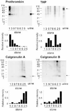
          Calcium oxalate kidney stones contain low amounts of proteins, some of which have been implicated in progression or prevention of kidney stone formation. To gain insights into the pathophysiology of urolithiasis, we have characterized protein components of calcium oxalate kidney stones by proteomic approaches. Proteins extracted from kidney stones showed highly heterogeneous migration patterns in gel electrophoresis as reported. This was likely to be mainly due to proteolytic degradation and protein-protein crosslinking of Tamm-Horsfall protein and prothrombin. Protein profiles of calcium oxalate kidney stones were obtained by in-solution protease digestion followed by nanoLC-MALDI-tandem mass spectrometry, which resulted in identification of a total of 92 proteins in stones from 9 urolithiasis patients. Further analysis showed that protein species and their relative amounts were highly variable among individual stones. Although proteins such as prothrombin, osteopontin, calgranulin A and calgranulin B were found in most stones tested, some samples had high contents of prothrombin and osteopontin, while others had high contents of calgranulins. In addition, calgranulin-rich stones had various neutrophil-enriched proteins such as myeloperoxidase and lactotransferrin. These proteomic profiles of individual kidney stones suggest that multiple systems composed of different groups of proteins including leucocyte-derived ones are differently involved in pathogenesis of individual kidney stones depending on situations.

          Related collections

          Most cited references29

          • Record: found
          • Abstract: found
          • Article: not found

          Neutrophil granules: a library of innate immunity proteins.

          Gene expression profiling has revealed that circulating neutrophils rest between two major bursts of transcriptional and protein synthetic activities. The first occurs in the bone marrow. This equips the neutrophil with stocks of innate defense armory that are packaged into different granule subsets. The second burst occurs when the neutrophil exits circulation and migrates into tissues to find, capture and phagocytose microorganisms. This burst results in the synthesis and secretion of cytokines and chemokines that support resolution of inflammation and healing of damaged tissue. Gene expression profiling has revealed that neutrophils express a variety of innate immunity proteins, known previously only to be expressed in other cells. Likewise, it has become clear that some proteins previously thought to be specific to the neutrophil are expressed in epithelial cells during inflammation.
            Bookmark
            • Record: found
            • Abstract: found
            • Article: not found

            Tamm-Horsfall protein is a critical renal defense factor protecting against calcium oxalate crystal formation.

            The tubular fluid of the mammalian kidney is often supersaturated with mineral salts, but crystallization rarely occurs under normal conditions. The unique ability of the kidney to avoid harmful crystal formation has long been attributed to the inhibitory activity of the urinary macromolecules, although few in vivo studies have been carried out to examine this hypothesis. Here we examined the role of Tamm-Horsfall protein (THP), the principal urinary protein, in urinary defense against renal calcium crystal formation, using a THP knockout model that we recently developed. Wild-type and THP knockout mice were examined for the spontaneous formation of renal calcium crystals using von Kossa staining. The susceptibility of these mice to experimentally induced renal crystal formation was evaluated by administering mice with ethylene glycol, a precursor of oxalate, and vitamin D(3), which increases calcium absorption. Renal calcium crystals were visualized by von Kossa stain, dark field microscopy with polarized light and scanning electron microscopy. Inactivating the THP gene in mouse embryonic stem cells results in spontaneous formation of calcium crystals in adult kidneys. Excessive intake of calcium and oxalate, precursors of the most common type of human renal stones, dramatically increases both the frequency and the severity of renal calcium crystal formation in THP-deficient, but not in wild-type mice. Under high calcium/oxalate conditions, the absence of THP triggers a marked, adaptive induction in renal epithelial cells of osteopontin (OPN), a potent inhibitor of bone mineralization and vascular calcification. Thus, OPN may serve as an inducible inhibitor of calcium crystallization, whereas THP can serve as a constitutive and apparently more effective inhibitor. These results provide the first in vivo evidence that THP is a critical urinary defense factor and suggest that its deficiency could be an important contributing factor in human nephrolithiasis, a condition afflicting tens of millions of people in the world annually.
              Bookmark
              • Record: found
              • Abstract: found
              • Article: not found

              Mechanism of calcium oxalate renal stone formation and renal tubular cell injury.

              Formation of calcium oxalate stones tends to increase with age and begins from the attachment of a crystal formed in the cavity of renal tubules to the surface of renal tubular epithelial cells. Though most of the crystals formed in the cavity of renal tubules are discharged as is in the urine, in healthy people, crystals that attach to the surface of renal tubular epithelial cells are thought to be digested by macrophages and/or lysosomes inside of cells. However, in individuals with hyperoxaluria or crystal urine, renal tubular cells are injured and crystals easily become attached to them. Various factors are thought to be involved in renal tubular cell injury. Crystals attached to the surface of renal tubular cells are taken into the cells (crystal-cell interaction). And then the crystal and crystal aggregates grow, and finally a stone is formed.
                Bookmark

                Author and article information

                Contributors
                Role: Editor
                Journal
                PLoS One
                PLoS ONE
                plos
                plosone
                PLoS ONE
                Public Library of Science (San Francisco, USA )
                1932-6203
                2013
                9 July 2013
                : 8
                : 7
                : e68624
                Affiliations
                [1 ]Laboratory of Homeostatic Integration, Division of Integrated Protein Functions, Institute for Protein Research, Osaka University, Suita, Osaka, Japan
                [2 ]Laboratory of Protein Profiling and Functional Proteomics, Research Center for Structural and Functional Proteomics, Institute for Protein Research, Osaka University, Suita, Osaka, Japan
                [3 ]Department of Urology, Osaka University Graduate School of Medicine, Suita, Osaka, Japan
                Federal University of São Paulo (UNIFESP), Escola Paulista de Medicina, Brazil
                Author notes

                Competing Interests: The authors have declared that no competing interests exist.

                Conceived and designed the experiments: MT NN AO TT. Performed the experiments: NO CM IY. Analyzed the data: NO KS TT. Wrote the paper: NO.

                [¤]

                Current address: Department of Urology, Osaka Rosai Hospital, Osaka, Japan

                Article
                PONE-D-13-05777
                10.1371/journal.pone.0068624
                3706363
                23874695
                6ba2e129-3412-43ca-a006-1424cfa5a29f
                Copyright @ 2013

                This is an open-access article distributed under the terms of the Creative Commons Attribution License, which permits unrestricted use, distribution, and reproduction in any medium, provided the original author and source are credited.

                History
                : 6 February 2013
                : 30 May 2013
                Page count
                Pages: 9
                Funding
                This work was supported by funds from the Ministry of Education, Culture, Sports, Science and Technology-Japan. The funders had no role in study design, data collection and analysis, decision to publish, or preparation of the manuscript. No additional external funding received for this study.
                Categories
                Research Article
                Biology
                Biochemistry
                Proteins
                Immune System Proteins
                Protein Classes
                Proteome
                Proteomics
                Protein Abundance
                Proteomic Databases
                Spectrometric Identification of Proteins
                Medicine
                Diagnostic Medicine
                Urology
                Kidney Stones

                Uncategorized
                Uncategorized

                Comments

                Comment on this article