10
views
0
recommends
+1 Recommend
0 collections
    0
    shares
      • Record: found
      • Abstract: found
      • Article: found
      Is Open Access

      Choroidal Thickness and microRNA146 in Lupus Nephritis Patients

      research-article

      Read this article at

      Bookmark
          There is no author summary for this article yet. Authors can add summaries to their articles on ScienceOpen to make them more accessible to a non-specialist audience.

          Abstract

          Purpose

          To evaluate the choroidal thickness (CT) in the macular area in patients with lupus nephritis and to compare the results with both non-nephritic patients and normal control. To assess the relation of CT to serum microRNA146, disease duration, activity index, and medications.

          Patients and Methods

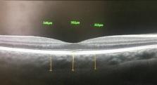
          Thirty-five SLE patients and thirty normal healthy controls were enrolled for this cross-sectional prospective study. All participants have undergone optical coherence tomography using RTVue OCT (Optovue Inc., Fremont, CA, USA). The scan used was the macular cross 6-mm line. We measured CT from the posterior edge of the retinal pigment epithelium (RPE) to the choroid-sclera junction at subfovea, and 750 µm both temporal and nasal to the fovea.

          Results

          The mean central subfoveal CT in patients was 275.7 ± 41.0 µm (214–374 µm), and the mean central subfoveal CT in the control group was 364.5± 23.0 µm (323–411µm). There was a significant thinning at all three points in patients compared to the control group (p<0.001, Mann–Whitney U-test). In the patients group, subfoveal choroid in non-nephritic subgroup showed significant thinning compared to nephritic subgroup (p=0.032, Mann–Whitney U-test). Drusen-like deposits (DLDs) were detected in 22.9% (8/35) of patients and none in control (p=.023). MiRNA146 showed a significant positive correlation with nephritic lupus patients (r=0.036, P=0.04).

          Conclusion

          The choroidal thickness was significantly thicker among the nephritic lupus patients as compared to the non-nephritic subgroup. Both SLE patients’ subgroups are thinner than normal control. Subfoveal choroidal thickening can be considered a biomarker in nephritic lupus especially in conjunction with an increase in miRNA146a. All SLE patients are at risk of small Drusen-like deposits.

          Related collections

          Most cited references24

          • Record: found
          • Abstract: found
          • Article: not found

          A pilot study of enhanced depth imaging optical coherence tomography of the choroid in normal eyes.

          To measure macular choroidal thickness in normal eyes at different points using enhanced depth imaging (EDI) optical coherence tomography (OCT) and to evaluate the association of choroidal thickness and age. Retrospective, observational case series. EDI OCT images were obtained in patients without significant retinal or choroidal pathologic features. The images were obtained by positioning a spectral-domain OCT device close enough to the eye to acquire an inverted image. Seven sections were obtained within a 5 x 30-degree area centered at the fovea, with 100 scans averaged for each section. The choroid was measured from the outer border of the retinal pigment epithelium to the inner scleral border at 500-microm intervals of a horizontal section from 3 mm temporal to the fovea to 3 mm nasal to the fovea. Statistical analysis was performed to evaluate variations of choroidal thickness at each location and to correlate choroidal thickness and patient age. The mean age of the 30 patients (54 eyes) was 50.4 years (range, 19 to 85 years), and 14 patients (46.7%) were female. The choroid was thickest underneath the fovea (mean, 287 microm; standard deviation, +/- 76 microm). Choroidal thickness decreased rapidly in the nasal direction and averaged 145 microm (+/- 57 microm) at 3 mm nasal to the fovea. Increasing age was correlated significantly with decreasing choroidal thickness at all points measured. Regression analysis suggested that the subfoveal choroidal thickness decreased by 15.6 microm for each decade of life. Choroidal thickness seems to vary topographically within the posterior pole. The thickness of the choroid showed a negative correlation with age. The decrease in the thickness of the choroid may play a role in the pathophysiologic features of various age-related ocular conditions.
            Bookmark
            • Record: found
            • Abstract: found
            • Article: not found

            MicroRNAs direct rapid deadenylation of mRNA.

            MicroRNAs (miRNAs) are ubiquitous regulators of eukaryotic gene expression. In addition to repressing translation, miRNAs can down-regulate the concentration of mRNAs that contain elements to which they are imperfectly complementary. Using miR-125b and let-7 as representative miRNAs, we show that in mammalian cells this reduction in message abundance is a consequence of accelerated deadenylation, which leads to rapid mRNA decay. The ability of miRNAs to expedite poly(A) removal does not result from decreased translation; nor does translational repression by miRNAs require a poly(A) tail, a 3' histone stem-loop being an effective substitute. These findings suggest that miRNAs use two distinct posttranscriptional mechanisms to down-regulate gene expression.
              Bookmark
              • Record: found
              • Abstract: found
              • Article: not found

              Choroidal thickness in normal eyes measured using Cirrus HD optical coherence tomography.

              To examine choroidal thickness and area in healthy eyes using spectral-domain optical coherence tomography (SD-OCT). Retrospective, observational case series. Thirty-four eyes (34 subjects), with no retinal or choroidal disease, underwent high-definition raster scanning using SD-OCT with frame enhancement software. Choroidal thickness was measured from the posterior edge of the retinal pigment epithelium to the choroid/sclera junction at 500-microm intervals up to 2500 microm temporal and nasal to the fovea. The central 1-mm area of the choroid was also measured, along with foveal thickness of the retina. All measurements were performed by 2 independent observers. Statistical analysis was used to correlate inter-observer findings, choroidal thickness and area measurements with age, and choroidal thickness with retinal foveal thickness. The 34 subjects had a mean age of 51.1 years. Reliable measurements of choroidal thickness were obtainable in 74% of eyes examined. Choroidal thickness and area measurements had strong inter-observer correlation (r = 0.92, P < .0001 and r = 0.93, P < .0001 respectively). Area had a moderate negative correlation with age (r = -0.62, P < .0001) that was comparable to the correlation between mean subfoveal choroidal thickness and age (r = -0.61, P < .0001). Retinal and choroidal thickness were found to be poorly correlated (r = -0.23, P = .18). Mean choroidal thickness showed a pattern of thinnest choroid nasally, thickening in the subfoveal region, and then thinning again temporally. Mean subfoveal choroidal thickness was found to be 272 microm (SD, +/- 81 microm). Choroidal thickness can be measured using SD-OCT high-definition raster scans in the majority of eyes. Choroidal thickness across the macula demonstrates a thin choroid nasally, thickest subfoveally, and again thinner temporally, and a trend toward decreasing choroidal thickness with age. Copyright (c) 2010 Elsevier Inc. All rights reserved.
                Bookmark

                Author and article information

                Journal
                Clin Ophthalmol
                Clin Ophthalmol
                OPTH
                clinop
                Clinical Ophthalmology (Auckland, N.Z.)
                Dove
                1177-5467
                1177-5483
                02 June 2020
                2020
                : 14
                : 1503-1510
                Affiliations
                [1 ]Ophthalmology Department, Kasr Alainy, Cairo University , Cairo, Egypt
                [2 ]Rheumatology and Rehabilitation, Kasr Alainy, Cairo University , Cairo, Egypt
                [3 ]Clinical Pathology, Kasr Alainy, Cairo University , Cairo, Egypt
                Author notes
                Correspondence: Shaymaa Hassan Salah Kasr Alainy Hospital, 1 Saray Al-Manial , Cairo11559, EgyptTel +201002229645Fax +20223636504 Email Shaimaa.Sayed@kasralainy.edu.eg
                Author information
                http://orcid.org/0000-0001-6381-6574
                http://orcid.org/0000-0002-5930-2885
                http://orcid.org/0000-0002-8602-3371
                Article
                254288
                10.2147/OPTH.S254288
                7276375
                32581507
                6bed2574-14bf-48d3-a44d-8e1c0430b541
                © 2020 Hassan Salah et al.

                This work is published and licensed by Dove Medical Press Limited. The full terms of this license are available at https://www.dovepress.com/terms.php and incorporate the Creative Commons Attribution – Non Commercial (unported, v3.0) License ( http://creativecommons.org/licenses/by-nc/3.0/). By accessing the work you hereby accept the Terms. Non-commercial uses of the work are permitted without any further permission from Dove Medical Press Limited, provided the work is properly attributed. For permission for commercial use of this work, please see paragraphs 4.2 and 5 of our Terms ( https://www.dovepress.com/terms.php).

                History
                : 17 March 2020
                : 11 May 2020
                Page count
                Figures: 5, Tables: 5, References: 33, Pages: 8
                Categories
                Original Research

                Ophthalmology & Optometry
                choroidal thinning,lupus nephritis,mirna146a,sledi
                Ophthalmology & Optometry
                choroidal thinning, lupus nephritis, mirna146a, sledi

                Comments

                Comment on this article