13
views
0
recommends
+1 Recommend
0 collections
    0
    shares
      • Record: found
      • Abstract: found
      • Article: found
      Is Open Access

      Concordance and Reproducibility of Melanoma Staging According to the 7th vs 8th Edition of the AJCC Cancer Staging Manual

      research-article

      Read this article at

      Bookmark
          There is no author summary for this article yet. Authors can add summaries to their articles on ScienceOpen to make them more accessible to a non-specialist audience.

          Abstract

          IMPORTANCE

          The recently updated American Joint Committee on Cancer (AJCC) classification of cancer staging, the AJCC Cancer Staging Manual , 8th edition ( AJCC 8), includes revisions to definitions of T1a vs T1b or greater. The Melanoma Pathology Study database affords a comparison,of pathologists’ concordance and reproducibility in the microstaging of melanoma according to both the existing 7th edition (AJCC 7) and the new AJCC 8 .

          OBJECTIVE

          To compare AJCC 7 and AJCC 8 to examine whether changes to the definitions of T1a and T1b or greater are associated with changes in concordance and reproducibility.

          DESIGN, SETTING, AND PARTICIPANTS

          In this diagnostic study conducted as part of the national Melanoma Pathology Study across US states, 187 pathologists interpreting melanocytic skin lesions in practice completed 4342 independent case interpretations of 116 invasive melanoma cases. A consensus reference diagnosis and participating pathologists’ interpretations were classified into the Melanocytic Pathology Assessment Tool and Hierarchy for Diagnosis class IV (T1a) or class V ( T1b) using both the AJCC 7 and AJCC 8 criteria.

          MAIN OUTCOMES AND MEASURES

          Concordance with consensus reference diagnosis, interobserver reproducibility, and intraobserver reproducibility.

          RESULTS

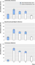
          For T1a diagnoses, participating pathologists’ concordance with the consensus reference diagnosis increased from 44% (95% CI, 41%−48%) to 54% (95% CI, 51%−57%) using AJCC 7 and AJCC 8 criteria, respectively. The concordance for cases of T1b or greater increased from 72% (95% CI, 69%−75%) to 78% (95% CI, 75%−80%). Intraobserver reproducibility of diagnoses also improved, increasing from 59% (95% CI, 56%−63%) to 64% (95% CI, 62%−67%) for T1a invasive melanoma, and from 74% (95% CI, 71%−76%) to 77% (95% CI, 74%−79%) for T1b or greater invasive melanoma cases.

          CONCLUSIONS AND RELEVANCE

          Melanoma staging in AJCC 8 shows greater reproducibility and higher concordance with a reference standard. Improved classification of invasive melanoma can be expected after implementation of AJCC 8, suggesting a positive impact on patients. However, despite improvement, concordance and reproducibility remain low.

          Related collections

          Most cited references13

          • Record: found
          • Abstract: found
          • Article: found
          Is Open Access

          Pathologists’ diagnosis of invasive melanoma and melanocytic proliferations: observer accuracy and reproducibility study

          Objective To quantify the accuracy and reproducibility of pathologists’ diagnoses of melanocytic skin lesions. Design Observer accuracy and reproducibility study. Setting 10 US states. Participants Skin biopsy cases (n=240), grouped into sets of 36 or 48. Pathologists from 10 US states were randomized to independently interpret the same set on two occasions (phases 1 and 2), at least eight months apart. Main outcome measures Pathologists’ interpretations were condensed into five classes: I (eg, nevus or mild atypia); II (eg, moderate atypia); III (eg, severe atypia or melanoma in situ); IV (eg, pathologic stage T1a (pT1a) early invasive melanoma); and V (eg, ≥pT1b invasive melanoma). Reproducibility was assessed by intraobserver and interobserver concordance rates, and accuracy by concordance with three reference diagnoses. Results In phase 1, 187 pathologists completed 8976 independent case interpretations resulting in an average of 10 (SD 4) different diagnostic terms applied to each case. Among pathologists interpreting the same cases in both phases, when pathologists diagnosed a case as class I or class V during phase 1, they gave the same diagnosis in phase 2 for the majority of cases (class I 76.7%; class V 82.6%). However, the intraobserver reproducibility was lower for cases interpreted as class II (35.2%), class III (59.5%), and class IV (63.2%). Average interobserver concordance rates were lower, but with similar trends. Accuracy using a consensus diagnosis of experienced pathologists as reference varied by class: I, 92% (95% confidence interval 90% to 94%); II, 25% (22% to 28%); III, 40% (37% to 44%); IV, 43% (39% to 46%); and V, 72% (69% to 75%). It is estimated that at a population level, 82.8% (81.0% to 84.5%) of melanocytic skin biopsy diagnoses would have their diagnosis verified if reviewed by a consensus reference panel of experienced pathologists, with 8.0% (6.2% to 9.9%) of cases overinterpreted by the initial pathologist and 9.2% (8.8% to 9.6%) underinterpreted. Conclusion Diagnoses spanning moderately dysplastic nevi to early stage invasive melanoma were neither reproducible nor accurate in this large study of pathologists in the USA. Efforts to improve clinical practice should include using a standardized classification system, acknowledging uncertainty in pathology reports, and developing tools such as molecular markers to support pathologists’ visual assessments.
            Bookmark
            • Record: found
            • Abstract: found
            • Article: not found

            Discordance in the histopathologic diagnosis of melanoma and melanocytic nevi between expert pathologists.

            The reliability of a diagnostic test depends on the reproducibility of the result. Many clinical diagnostic tests can be quantified with established ranges and standard deviations. Other tests are more subjective, such as those that depend on analysis of a visual image with an increased possibility of variance in the result. To study this variance, the authors analyzed the performance of expert pathologists in the interpretation of cutaneous melanocytic tumors. A panel of expert pathologists was convened to review anatomic pathology specimens from melanocytic tumors. Each pathologist submitted five specimens, from which 37 were selected for review. Only one slide was used for each case. All specimens were interpreted by each pathologist without consultation with each other. In addition to standard diagnostic terms, each specimen was designated as benign, malignant, or indeterminate. Statistical analysis was used to determine the degree of concordance. The combined kappa statistic for the eight observers and three possible outcomes (benign, malignant, or indeterminate) was 0.50. A kappa statistic of this magnitude, is defined as being moderate. In 62% of the specimens, there was unanimous agreement or only one discordant designation. Thirty-eight percent had two or more discordant interpretations. No single pathologist had a disproportionate number of discordant designations. This study mimics the consultation practice of anatomic pathology and shows the variability and discordance in diagnostic language and designation of biological behavior. The results suggest the criteria for the diagnosis of melanomas and melanocytic nevi need to be refined and more consistently applied.
              Bookmark
              • Record: found
              • Abstract: found
              • Article: not found

              The MPATH-Dx reporting schema for melanocytic proliferations and melanoma

              The histologic diagnosis of melanoma and nevi can be subject to discordance and errors, potentially leading to inappropriate treatment and harm. Diagnostic terminology is not standardized, creating confusion for providers and patients and challenges for investigators.
                Bookmark

                Author and article information

                Journal
                101729235
                47705
                JAMA Netw Open
                JAMA Netw Open
                JAMA network open
                2574-3805
                23 June 2018
                May 2018
                14 December 2018
                : 1
                : 1
                : e180083
                Affiliations
                Department of Medicine, David Geffen School of Medicine, University of California, Los Angeles (Elmore); Department of Pathology and Laboratory Medicine, Hospital of the University of Pennsylvania, Philadelphia (Elder); Department of Pathology, Institut Curie, Paris, France (Barnhill); Paris Sciences and Lettres Research University, Paris, France (Barnhill); Faculty of Medicine, University of Paris Descartes, Paris, France (Barnhill); Pathology Associates, Clovis, California (Knezevich); Program in Biostatistics and Biomathematics, Fred Hutchinson Cancer Research Center, Seattle, Washington (Longton, Pepe); Department of Epidemiology, Geisel School of Medicine at Dartmouth, Hanover, New Hampshire (Titus); Department of Pediatrics, Geisel School of Medicine at Dartmouth, Hanover, New Hampshire (Titus); Norris Cotton Cancer Center, Lebanon, New Hampshire (Titus); Center for Dermatoepidemiology, Providence Veterans Affair Medical Center, Providence, Rhode Island (Weinstock); Department of Dermatology, Brown University, Providence, Rhode Island (Weinstock); Department of Epidemiology, Brown University, Providence, Rhode Island (Weinstock); Department of Medical Informatics and Clinical Epidemiology, School of Medicine, Oregon Health and Science University, Portland (Nelson); Department of Medicine, School of Medicine, Oregon Health and Science University, Portland (Nelson); Department of Medicine, University of Washington School of Medicine, Seattle (Reisch, Radick, Piepkorn); Dermatopathology Northwest, Bellevue, Washington (Piepkorn)
                Author notes
                Corresponding Author: Joann G. Elmore, MD, MPH, Department of Medicine, David Geffen School of Medicine, University of California, Los Angeles, 10940 Wilshire Blvd, Ste 710, Los Angeles, CA 90024 ( jelmore@ 123456mednet.ucl.edu ).
                Article
                NIHMS976104
                10.1001/jamanetworkopen.2018.0083
                6294444
                6de5e332-1b1a-4cd3-bc66-c4579a39a25e

                Open Access: This is an open access article distributed under the terms of the CC002DBY License.

                History
                Categories
                Article

                Comments

                Comment on this article