1
views
0
recommends
+1 Recommend
0 collections
    0
    shares
      • Record: found
      • Abstract: found
      • Article: found
      Is Open Access

      Keratin-mediated hair growth and its underlying biological mechanism

      research-article

      Read this article at

      Bookmark
          There is no author summary for this article yet. Authors can add summaries to their articles on ScienceOpen to make them more accessible to a non-specialist audience.

          Abstract

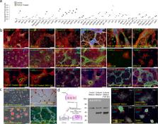
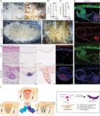
          Here we show that intradermal injection of keratin promotes hair growth in mice, which results from extracellular interaction of keratin with hair forming cells. Extracellular application of keratin induces condensation of dermal papilla cells and the generation of a P-cadherin-expressing cell population (hair germ) from outer root sheath cells via keratin-mediated microenvironmental changes. Exogenous keratin-mediated hair growth is reflected by the finding that keratin exposure from transforming growth factor beta 2 (TGFβ2)-induced apoptotic outer root sheath cells appears to be critical for dermal papilla cell condensation and P-cadherin-expressing hair germ formation. Immunodepletion or downregulation of keratin released from or expressed in TGFβ2-induced apoptotic outer root sheath cells negatively influences dermal papilla cell condensation and hair germ formation. Our pilot study provides an evidence on initiating hair regeneration and insight into the biological function of keratin exposed from apoptotic epithelial cells in tissue regeneration and development.

          Abstract

          Injecting human hair-derived keratin into mice skin accelerates hair growth & formation, as TGFβ2 secretion during hair destruction stimulates epithelial cell death and keratin release, leading to dermal cell condensation & hair growth.

          Related collections

          Most cited references50

          • Record: found
          • Abstract: found
          • Article: not found

          Defining the epithelial stem cell niche in skin.

          Many adult regenerative cells divide infrequently but have high proliferative capacity. We developed a strategy to fluorescently label slow-cycling cells in a cell type-specific fashion. We used this method to purify the label-retaining cells (LRCs) that mark the skin stem cell (SC) niche. We found that these cells rarely divide within their niche but change properties abruptly when stimulated to exit. We determined their transcriptional profile, which, when compared to progeny and other SCs, defines the niche. Many of the >100 messenger RNAs preferentially expressed in the niche encode surface receptors and secreted proteins, enabling LRCs to signal and respond to their environment.
            Bookmark
            • Record: found
            • Abstract: not found
            • Article: not found

            β-Catenin Controls Hair Follicle Morphogenesis and Stem Cell Differentiation in the Skin

              Bookmark
              • Record: found
              • Abstract: found
              • Article: found
              Is Open Access

              Hedgehog stimulates hair follicle neogenesis by creating inductive dermis during murine skin wound healing

              Mammalian wounds typically heal by fibrotic repair without hair follicle (HF) regeneration. Fibrosis and regeneration are currently considered the opposite end of wound healing. This study sought to determine if scar could be remodeled to promote healing with HF regeneration. Here, we identify that activation of the Sonic hedgehog (Shh) pathway reinstalls a regenerative dermal niche, called dermal papilla, which is required and sufficient for HF neogenesis (HFN). Epidermal Shh overexpression or constitutive Smoothened dermal activation results in extensive HFN in wounds that otherwise end in scarring. While long-term Wnt activation is associated with fibrosis, Shh signal activation in Wnt active cells promotes the dermal papilla fate in scarring wounds. These studies demonstrate that mechanisms of scarring and regeneration are not distant from one another and that wound repair can be redirected to promote regeneration following injury by modifying a key dermal signal.
                Bookmark

                Author and article information

                Contributors
                yshwang@khu.ac.kr
                Journal
                Commun Biol
                Commun Biol
                Communications Biology
                Nature Publishing Group UK (London )
                2399-3642
                19 November 2022
                19 November 2022
                2022
                : 5
                : 1270
                Affiliations
                [1 ]GRID grid.289247.2, ISNI 0000 0001 2171 7818, Department of Maxillofacial Biomedical Engineering, College of Dentistry, , Kyung Hee University, ; Seoul, 02447 Republic of Korea
                [2 ]GRID grid.258676.8, ISNI 0000 0004 0532 8339, Department of Veterinary Clinical Pathology, College of Veterinary Medicine, , Konkuk University, ; 120 Neungdong-ro, Gwangjin-gu, Seoul, 05029 Republic of Korea
                [3 ]GRID grid.289247.2, ISNI 0000 0001 2171 7818, Department of Oral Microbiology, College of Dentistry, , Kyung Hee University, ; Seoul, 02447 Republic of Korea
                [4 ]KeraMedix Inc, # 204, Open Innovation Bld, Hongryeung Bio-Cluster, 117-3 Hoegi-ro, Dongdaemun-gu, Seoul, 02455 Republic of Korea
                [5 ]GRID grid.289247.2, ISNI 0000 0001 2171 7818, Department of Dental Materials, College of Dentistry, , Kyung Hee University, ; Seoul, 02447 Republic of Korea
                [6 ]Headquarters of New Drug Development Support, Chemon Inc. 15 F, Gyeonggi Bio Center, Cheongju, Gyeonggi-do 16229 Republic of Korea
                [7 ]GRID grid.411311.7, ISNI 0000 0004 0532 4733, Present Address: Department of Dental Hygiene, College of Health Science, , Cheongju University, ; Cheongju, 360-764 Republic of Korea
                [8 ]GRID grid.419901.4, Present Address: Terasaki Institute for Biomedical Innovation, ; Los Angeles, CA 90064 USA
                Author information
                http://orcid.org/0000-0002-5085-6988
                http://orcid.org/0000-0003-2372-5269
                http://orcid.org/0000-0001-6197-7542
                Article
                4232
                10.1038/s42003-022-04232-9
                9675858
                36402892
                6e9b75c8-493b-4ca4-92ac-bb2014210cbd
                © The Author(s) 2022

                Open Access This article is licensed under a Creative Commons Attribution 4.0 International License, which permits use, sharing, adaptation, distribution and reproduction in any medium or format, as long as you give appropriate credit to the original author(s) and the source, provide a link to the Creative Commons license, and indicate if changes were made. The images or other third party material in this article are included in the article’s Creative Commons license, unless indicated otherwise in a credit line to the material. If material is not included in the article’s Creative Commons license and your intended use is not permitted by statutory regulation or exceeds the permitted use, you will need to obtain permission directly from the copyright holder. To view a copy of this license, visit http://creativecommons.org/licenses/by/4.0/.

                History
                : 1 November 2020
                : 8 November 2022
                Categories
                Article
                Custom metadata
                © The Author(s) 2022

                biomaterials - cells,stem-cell niche
                biomaterials - cells, stem-cell niche

                Comments

                Comment on this article