44
views
0
recommends
+1 Recommend
0 collections
    0
    shares
      • Record: found
      • Abstract: found
      • Article: found
      Is Open Access

      Predisposing Factors, Microbial Characteristics, and Clinical Outcome of Microbial Keratitis in a Tertiary Centre in Hong Kong: A 10-Year Experience

      research-article

      Read this article at

      Bookmark
          There is no author summary for this article yet. Authors can add summaries to their articles on ScienceOpen to make them more accessible to a non-specialist audience.

          Abstract

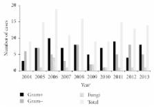
          Purpose. To study the risk factors, microbial profile, antibiotic susceptibility pattern, and outcome for microbial keratitis over the past 10 years in a tertiary center in Hong Kong. Methods. All cases with corneal scraping performed in Queen Mary Hospital, Hong Kong from January 2004 to December 2013 were included. Clinical outcome was defined as poor if the final visual acuity (VA) was abnormal or worse than presenting VA, a major complication occurred, or therapeutic keratoplasty was required. Results. 347 scrapes were performed in the 10-year period growing 130 microorganisms (32.3% culture positive rate). Contact lens use was the commonest risk factor. The commonest isolates were coagulase-negative Staphylococcus and Pseudomonas aeruginosa. Fluoroquinolone susceptibility was tested in 47 Gram-negative bacteria with 93.6% susceptibility (100% for Pseudomonas). 90.7% of cases had good visual outcome. Multivariate logistic regression showed age ( p = 0.03), trauma ( p = 0.006), and ulcer size >3 mm ( p = 0.039) to be independently associated with poor outcome. Conclusion. There was no shifting trend in the isolate distribution or emergence of resistant strains in our study. Contact lens wear was the commonest risk factor, with Pseudomonas being the most frequent isolate in this group. It remained 100% susceptible to fluoroquinolones and 97% cases had good visual outcome.

          Related collections

          Most cited references25

          • Record: found
          • Abstract: found
          • Article: not found

          Bacterial keratitis: predisposing factors, clinical and microbiological review of 300 cases.

          To identify predisposing factors and to define clinical and microbiological characteristics of bacterial keratitis in current practice. A retrospective analysis of the hospital records of patients presenting with bacterial keratitis and treated at the Quinze-Vingts National Center of Ophthalmology, Paris, France, was performed during a 20 month period. A bacterial keratitis was defined as a suppurative corneal infiltrate and overlying epithelial defect associated with presence of bacteria on corneal scraping and/or that was cured with antibiotic therapy. Risk factors, clinical and microbiological data were collected. 300 cases (291 patients) of presumed bacterial keratitis were included. Potential predisposing factors, usually multiple, were identified in 90.6% of cases. Contact lens wear was the main risk factor (50.3%). Trauma or a history of keratopathy was found in 15% and 21% of cases, respectively. An organism was identified in 201 eyes (68%). 83% of the infections involved Gram positive bacteria, 17% involved Gram negative bacteria, and 2% were polymicrobial. Gram negative bacteria were associated with severe anterior chamber inflammation (p=0.004), as well as greater surface of infiltrates (p=0.01). 99% of ulcers resolved with treatment, but only 60% of patients had visual acuity better than the level at admission, and 5% had very poor visual outcome. Contact lens wear is the most important risk factor. Most community acquired bacterial ulcers resolve with appropriate treatment.
            Bookmark
            • Record: found
            • Abstract: found
            • Article: not found

            Risk factors and causative organisms in microbial keratitis.

            To establish the risk factors, causative organisms, levels of antibiotic resistance, patient demographics, clinical presentations, and clinical outcomes of microbial keratitis at a tertiary hospital in Australia. Patients who had a corneal scraping for culture over a 5-year period were identified through the local microbiology database, and a retrospective audit of their medical records was carried out. Clinical information was gathered from medical records, and smear, culture, and antibiotic resistance results were from the microbiology database. An index of disease severity was calculated for each patient from scores for the magnitude of the epithelial defect and anterior-chamber reaction and the location of the lesion. Associations between risk factors for keratitis and variables such as patient demographics, causative organism and antibiotic resistance, disease severity, and outcome were analyzed by using analysis of variance and chi tests with appropriate correction for multiple comparisons. Two hundred fifty-three cases of microbial keratitis in 231 patients were included. Sixty percent of patients were men, and there was a bimodal distribution in the age of presentation. Common risk factors for keratitis were contact lens wear (53; 22%), ocular surface disease (45; 18%), ocular trauma (41; 16%), and prior ocular surgery (28; 11%). Gram stains were positive in 33%, with a sensitivity of 53% and specificity of 89%. Cultures of corneal scrapings were positive in 65% of cases, and Pseudomonas aeruginosa (44; 17%), coagulase-negative staphylococci (22; 9%), Staphylococcus aureus (19; 8%), and fungi (7; 3%) were commonly recovered. P. aeruginosa was more common than other culture results in contact lens-related cases (55% vs. 0%-23%; P < 0.001), and S. aureus was more common than other culture results in ocular surgery-related cases (29% vs. 0%-21%; P < 0.001). Patients with keratitis related to prior ocular surface disease had more severe keratitis at the time of scraping (P = 0.037). Cultures positive for Fusarium, P. aeruginosa, and other Gram-negative organisms had statistically significantly more severe keratitis at the time of scraping, whereas patients with negative cultures had milder keratitis (P = 0.030). Only 2% of all bacterial isolates were resistant to ciprofloxacin, 20% of Gram-positive isolates were resistant to cephalothin, and no Gram-negative isolates were resistant to gentamicin. In this series, the most common risk factor for keratitis was contact lens wear and the most commonly isolated organism was P. aeruginosa.
              Bookmark
              • Record: found
              • Abstract: found
              • Article: found
              Is Open Access

              Review of epidemiological features, microbiological diagnosis and treatment outcome of microbial keratitis: Experience of over a decade

              Purpose: To review the epidemiological characteristics, microbiological profile, and treatment outcome of patients with suspected microbial keratitis. Materials and Methods: Retrospective analysis of a non-comparative series from the database was done. All the patients presenting with corneal stromal infiltrate underwent standard microbiologic evaluation of their corneal scrapings, and smear and culture-guided antimicrobial therapy. Results: Out of 5897 suspected cases of microbial keratitis 3563 (60.4%) were culture-proven (bacterial – 1849, 51.9%; fungal – 1360, 38.2%; Acanthamoeba – 86, 2.4%; mixed – 268, 7.5%). Patients with agriculture-based activities were at 1.33 times (CI 1.16–1.51) greater risk of developing microbial keratitis and patients with ocular trauma were 5.33 times (CI 6.41–6.44) more likely to develop microbial keratitis. Potassium hydroxide with calcofluor white was most sensitive for detecting fungi (90.6%) and Acanthamoeba (84.0%) in corneal scrapings, however, Gram stain had a low sensitivity of 56.6% in detection of bacteria. Majority of the bacterial infections were caused by Staphylococcus epidermidis (42.3%) and Fusarium species (36.6%) was the leading cause of fungal infections. A significantly larger number of patients (691/1360, 50.8%) with fungal keratitis required surgical intervention compared to bacterial (799/1849, 43.2%) and Acanthamoeba (15/86, 17.4%) keratitis. Corneal healed scar was achieved in 75.5%, 64.8%, and 90.0% of patients with bacterial, fungal, and Acanthamoeba keratitis respectively. Conclusions: While diagnostic and treatment modalities are well in place the final outcome is suboptimal in fungal keratitis. With more effective treatment available for bacterial and Acanthamoeba keratitis, the treatment of fungal keratitis is truly a challenge.
                Bookmark

                Author and article information

                Journal
                J Ophthalmol
                J Ophthalmol
                JOPH
                Journal of Ophthalmology
                Hindawi Publishing Corporation
                2090-004X
                2090-0058
                2015
                18 June 2015
                : 2015
                : 769436
                Affiliations
                1Department of Ophthalmology, LKS Faculty of Medicine, The University of Hong Kong, Pok Fu Lam, Hong Kong
                2Department of Microbiology, LKS Faculty of Medicine, The University of Hong Kong, Pok Fu Lam, Hong Kong
                3Department of Ophthalmology, Queen Mary Hospital, Pok Fu Lam, Hong Kong
                Author notes
                *Ian Yat-Hin Wong: ianyhwong@ 123456gmail.com

                Academic Editor: Francisco Arnalich-Montiel

                Article
                10.1155/2015/769436
                4488544
                26167295
                6f9c7a43-159b-4f63-a597-8ddc0d911144
                Copyright © 2015 Alex Lap-Ki Ng et al.

                This is an open access article distributed under the Creative Commons Attribution License, which permits unrestricted use, distribution, and reproduction in any medium, provided the original work is properly cited.

                History
                : 22 March 2015
                : 10 June 2015
                Categories
                Research Article

                Ophthalmology & Optometry
                Ophthalmology & Optometry

                Comments

                Comment on this article