8
views
0
recommends
+1 Recommend
0 collections
    0
    shares
      • Record: found
      • Abstract: found
      • Article: found
      Is Open Access

      Cannabinoids and Cannabinoid Receptors: The Story so Far

      review-article

      Read this article at

      Bookmark
          There is no author summary for this article yet. Authors can add summaries to their articles on ScienceOpen to make them more accessible to a non-specialist audience.

          Abstract

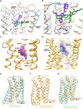
          Like most modern molecular biology and natural product chemistry, understanding cannabinoid pharmacology centers around molecular interactions, in this case, between the cannabinoids and their putative targets, the G-protein coupled receptors (GPCRs) cannabinoid receptor 1 (CB 1) and cannabinoid receptor 2 (CB 2). Understanding the complex structure and interplay between the partners in this molecular dance is required to understand the mechanism of action of synthetic, endogenous, and phytochemical cannabinoids. This review, with 91 references, surveys our understanding of the structural biology of the cannabinoids and their target receptors including both a critical comparison of the extant crystal structures and the computationally derived homology models, as well as an in-depth discussion about the binding modes of the major cannabinoids. The aim is to assist in situating structural biochemists, synthetic chemists, and molecular biologists who are new to the field of cannabis research.

          Graphical Abstract

          Highlights

          • Cannabinoid research has greatly expanded

          • Structural biology and computational chemistry jointly provide mechanistic insight

          • Structural data are being generated at an exponentially increasing rate

          • Phytocannabinoid targeting of other GPCR receptors deserves investigation

          Abstract

          Medical Substance; Supramolecular Chemistry; Molecular Biology; Structural Biology

          Related collections

          Most cited references73

          • Record: found
          • Abstract: found
          • Article: not found

          Molecular characterization of a peripheral receptor for cannabinoids.

          The major active ingredient of marijuana, delta 9-tetrahydrocannabinol (delta 9-THC), has been used as a psychoactive agent for thousands of years. Marijuana, and delta 9-THC, also exert a wide range of other effects including analgesia, anti-inflammation, immunosuppression, anticonvulsion, alleviation of intraocular pressure in glaucoma, and attenuation of vomiting. The clinical application of cannabinoids has, however, been limited by their psychoactive effects, and this has led to interest in the biochemical bases of their action. Progress stemmed initially from the synthesis of potent derivatives of delta 9-THC, and more recently from the cloning of a gene encoding a G-protein-coupled receptor for cannabinoids. This receptor is expressed in the brain but not in the periphery, except for a low level in testes. It has been proposed that the nonpsychoactive effects of cannabinoids are either mediated centrally or through direct interaction with other, non-receptor proteins. Here we report the cloning of a receptor for cannabinoids that is not expressed in the brain but rather in macrophages in the marginal zone of spleen.
            Bookmark
            • Record: found
            • Abstract: found
            • Article: not found

            High-resolution crystal structure of an engineered human beta2-adrenergic G protein-coupled receptor.

            Heterotrimeric guanine nucleotide-binding protein (G protein)-coupled receptors constitute the largest family of eukaryotic signal transduction proteins that communicate across the membrane. We report the crystal structure of a human beta2-adrenergic receptor-T4 lysozyme fusion protein bound to the partial inverse agonist carazolol at 2.4 angstrom resolution. The structure provides a high-resolution view of a human G protein-coupled receptor bound to a diffusible ligand. Ligand-binding site accessibility is enabled by the second extracellular loop, which is held out of the binding cavity by a pair of closely spaced disulfide bridges and a short helical segment within the loop. Cholesterol, a necessary component for crystallization, mediates an intriguing parallel association of receptor molecules in the crystal lattice. Although the location of carazolol in the beta2-adrenergic receptor is very similar to that of retinal in rhodopsin, structural differences in the ligand-binding site and other regions highlight the challenges in using rhodopsin as a template model for this large receptor family.
              Bookmark
              • Record: found
              • Abstract: found
              • Article: not found

              Crystal structure of rhodopsin: A G protein-coupled receptor.

              Heterotrimeric guanine nucleotide-binding protein (G protein)-coupled receptors (GPCRs) respond to a variety of different external stimuli and activate G proteins. GPCRs share many structural features, including a bundle of seven transmembrane alpha helices connected by six loops of varying lengths. We determined the structure of rhodopsin from diffraction data extending to 2.8 angstroms resolution. The highly organized structure in the extracellular region, including a conserved disulfide bridge, forms a basis for the arrangement of the seven-helix transmembrane motif. The ground-state chromophore, 11-cis-retinal, holds the transmembrane region of the protein in the inactive conformation. Interactions of the chromophore with a cluster of key residues determine the wavelength of the maximum absorption. Changes in these interactions among rhodopsins facilitate color discrimination. Identification of a set of residues that mediate interactions between the transmembrane helices and the cytoplasmic surface, where G-protein activation occurs, also suggests a possible structural change upon photoactivation.
                Bookmark

                Author and article information

                Contributors
                Journal
                iScience
                iScience
                iScience
                Elsevier
                2589-0042
                20 June 2020
                24 July 2020
                20 June 2020
                : 23
                : 7
                : 101301
                Affiliations
                [1 ]Department of Chemistry and Biochemistry, University of Windsor, Windsor, ON N9B 3P4, Canada
                Author notes
                []Corresponding author j.trant@ 123456uwindsor.ca
                Article
                S2589-0042(20)30488-0 101301
                10.1016/j.isci.2020.101301
                7339067
                32629422
                70a735cd-7b7f-4a91-8579-d8a16b452e4e
                © 2020 The Author(s)

                This is an open access article under the CC BY license (http://creativecommons.org/licenses/by/4.0/).

                History
                Categories
                Review

                medical substance,supramolecular chemistry,molecular biology,structural biology

                Comments

                Comment on this article