19
views
0
recommends
+1 Recommend
0 collections
    0
    shares
      • Record: found
      • Abstract: found
      • Article: found
      Is Open Access

      Targeting light-gated chloride channels to neuronal somatodendritic domain reduces their excitatory effect in the axon

      research-article

      Read this article at

      Bookmark
          There is no author summary for this article yet. Authors can add summaries to their articles on ScienceOpen to make them more accessible to a non-specialist audience.

          Abstract

          Light-gated chloride channels are emerging as promising optogenetic tools for inhibition of neural activity. However, their effects depend on the transmembrane chloride electrochemical gradient and may be complex due to the heterogeneity of this gradient in different developmental stages, neuronal types, and subcellular compartments. Here we characterized a light-gated chloride channel, GtACR2, in mouse cortical neurons. We found that GtACR2 activation inhibited the soma, but unexpectedly depolarized the presynaptic terminals resulting in neurotransmitter release. Other light-gated chloride channels had similar effects. Reducing the chloride concentrations in the axon and presynaptic terminals diminished the GtACR2-induced neurotransmitter release, indicating an excitatory effect of chloride channels in these compartments. A novel hybrid somatodendritic targeting motif reduced the GtACR2-induced neurotransmitter release while enhancing the somatic photocurrents. Our results highlight the necessity of precisely determining the effects of light-gated chloride channels under specific experimental conditions and provide a much-improved light-gated chloride channel for optogenetic inhibition.

          eLife digest

          One way to study the role of a specific neuron is to activate or inhibit the cell and then observe the consequences. This can be achieved by using optogenetics, a technique that involves introducing ‘light-gated’ ion channels in the outer membrane of a target neuron. When light is shone on the cell, these pore-like proteins open their channels: this allows ions to move into or out of the neuron.

          Ions flow from high concentration to low concentration areas. Typically, when a neuron is at rest, there are fewer chloride ions inside the cell than outside. Activating a light-gated chloride channel should thus cause these negatively charged ions to enter the neuron. The charge inside of the cell would become more negative relative to the outside: this would inhibit the neuron, making it less likely to fire.

          Here, Messier et al. looked into using a light-gated chlorine channel called GtACR2 to inhibit the activity of neurons in mouse brain slices, but the results were not as expected. Activating the chloride channel did inhibit the cell body, the area of the neuron that contains the nucleus. Yet, it had the opposite effect in the axon, the structure that carries electrical signals away from the cell body.

          There, activating GtACR2 caused chloride ions to leave the axon, which resulted in the neuron firing. Testing other types of optogenetic chloride channels produced the same result. Further experiments revealed that the concentration of chloride ions is higher inside the axon than the cell body, explaining the observed effects.

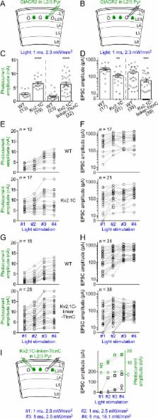
          Messier et al. then tried to redistribute the channels from the axon to the cell body, where the proteins are inhibitory. This was accomplished by fitting GtACR2 with a molecular tag that acts like an address label, with the cell body as the target destination. Overall, when these modified channels were activated, the neuron was more strongly inhibited.

          Ultimately, the GtACR2 channel designed by Messier et al. is a powerful new inhibitory optogenetic tool. In addition, this tool could be used to study chloride gradients in brain regions, cell types and areas of cells that are otherwise difficult to access.

          Related collections

          Most cited references39

          • Record: found
          • Abstract: found
          • Article: not found

          Transgenic mice for intersectional targeting of neural sensors and effectors with high specificity and performance.

          An increasingly powerful approach for studying brain circuits relies on targeting genetically encoded sensors and effectors to specific cell types. However, current approaches for this are still limited in functionality and specificity. Here we utilize several intersectional strategies to generate multiple transgenic mouse lines expressing high levels of novel genetic tools with high specificity. We developed driver and double reporter mouse lines and viral vectors using the Cre/Flp and Cre/Dre double recombinase systems and established a new, retargetable genomic locus, TIGRE, which allowed the generation of a large set of Cre/tTA-dependent reporter lines expressing fluorescent proteins, genetically encoded calcium, voltage, or glutamate indicators, and optogenetic effectors, all at substantially higher levels than before. High functionality was shown in example mouse lines for GCaMP6, YCX2.60, VSFP Butterfly 1.2, and Jaws. These novel transgenic lines greatly expand the ability to monitor and manipulate neuronal activities with increased specificity.
            Bookmark
            • Record: found
            • Abstract: found
            • Article: not found

            High-Performance Genetically Targetable Optical Neural Silencing via Light-Driven Proton Pumps

            The ability to silence the activity of genetically specified neurons in a temporally precise fashion would open up the ability to investigate the causal role of specific cell classes in neural computations, behaviors, and pathologies. Here we show that members of the class of light-driven outward proton pumps can mediate very powerful, safe, multiple-color silencing of neural activity. The gene archaerhodopsin-31 (Arch) from Halorubrum sodomense enables near-100% silencing of neurons in the awake brain when virally expressed in mouse cortex and illuminated with yellow light. Arch mediates currents of several hundred picoamps at low light powers, and supports neural silencing currents approaching 900 pA at light powers easily achievable in vivo. In addition, Arch spontaneously recovers from light-dependent inactivation, unlike light-driven chloride pumps that enter long-lasting inactive states in response to light. These properties of Arch are appropriate to mediate the optical silencing of significant brain volumes over behaviourally-relevant timescales. Arch function in neurons is well tolerated because pH excursions created by Arch illumination are minimized by self-limiting mechanisms to levels comparable to those mediated by channelrhodopsins2,3 or natural spike firing. To highlight how proton pump ecological and genomic diversity may support new innovation, we show that the blue-green light-drivable proton pump from the fungus Leptosphaeria maculans 4 (Mac) can, when expressed in neurons, enable neural silencing by blue light, thus enabling alongside other developed reagents the potential for independent silencing of two neural populations by blue vs. red light. Light-driven proton pumps thus represent a high-performance and extremely versatile class of “optogenetic” voltage and ion modulator, which will broadly empower new neuroscientific, biological, neurological, and psychiatric investigations.
              Bookmark
              • Record: found
              • Abstract: found
              • Article: not found

              ReaChR: A red-shifted variant of channelrhodopsin enables deep transcranial optogenetic excitation

              Channelrhodopsins are used to optogenetically depolarize neurons. We engineered a variant of channelrhodopsin, denoted Re d- a ctivatable Ch annel r hodopsin (ReaChR), that is optimally excited with orange to red light (λ ~ 590 to 630 nm) and offers improved membrane trafficking, higher photocurrents, and faster kinetics compared with existing red-shifted channelrhodopsins. Red light is more weakly scattered by tissue and absorbed less by blood than the blue to green wavelengths required by other channelrhodopsin variants. ReaChR expressed in vibrissa motor cortex was used to drive spiking and vibrissa motion in awake mice when excited with red light through intact skull. Precise vibrissa movements were evoked by expressing ReaChR in the facial motor nucleus in the brainstem and illuminating with red light through the external auditory canal. Thus, ReaChR enables transcranial optical activation of neurons in deep brain structures without the need to surgically thin the skull, form a transcranial window, or implant optical fibers.
                Bookmark

                Author and article information

                Contributors
                Role: Reviewing Editor
                Role: Senior Editor
                Journal
                eLife
                Elife
                eLife
                eLife
                eLife Sciences Publications, Ltd
                2050-084X
                09 August 2018
                2018
                : 7
                : e38506
                Affiliations
                [1 ]deptDepartment of Neuroscience Baylor College of Medicine Houston, TexasUnited States
                [2 ]deptThe Cain Foundation Laboratories Jan and Dan Duncan Neurological Research Institute at Texas Children’s Hospital Houston, TexasUnited States
                [3 ]deptDepartment of Molecular and Human Genetics Baylor College of Medicine Houston, TexasUnited States
                Northwestern University United States
                Vollum Institute United States
                Northwestern University United States
                Author information
                http://orcid.org/0000-0002-5865-7043
                https://orcid.org/0000-0003-4034-2884
                http://orcid.org/0000-0003-1463-8884
                Article
                38506
                10.7554/eLife.38506
                6130974
                30091701
                71f5d1ca-413c-4826-8fb5-79d4ee4161cb
                © 2018, Messier et al

                This article is distributed under the terms of the Creative Commons Attribution License, which permits unrestricted use and redistribution provided that the original author and source are credited.

                History
                : 19 May 2018
                : 08 August 2018
                Funding
                Funded by: FundRef http://dx.doi.org/10.13039/100001391, Whitehall Foundation;
                Award ID: 2015-05-54
                Award Recipient :
                Funded by: FundRef http://dx.doi.org/10.13039/100001234, Robert and Janice McNair Foundation;
                Award ID: MD/PhD Student Scholarship
                Award Recipient :
                Funded by: FundRef http://dx.doi.org/10.13039/100000002, National Institutes of Health;
                Award ID: R01NS100893
                Award Recipient :
                The funders had no role in study design, data collection and interpretation, or the decision to submit the work for publication.
                Categories
                Research Article
                Neuroscience
                Custom metadata
                A high axonal chloride concentration explains why activation of light-gated chloride channels causes neurotransmitter release, and a novel hybrid somatodendritic targeting motif ameliorates this phenomenon and improves their inhibitory function.

                Life sciences
                telencephalin,potassium channel kv2.1,axonal chloride,optogenetic inhibition,ic++,ichloc,mouse

                Comments

                Comment on this article